Saber-toothed cats were fierce and family-oriented

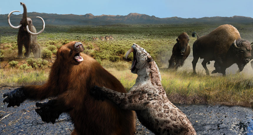
The adolescent saber-toothed cat on a summertime hunt realized too late that she had made a terrible miscalculation.

Already the size of a modern-day tiger, with huge canine teeth, she had crept across grassy terrain to ambush a giant ground sloth bellowing in distress. Ready to pounce, the cat’s front paw sank into sticky ground. Pressing down with her other three paws to free herself, then struggling in what has been called “tar pit aerobics,” she became irrevocably mired alongside her prey.

Scenarios much like this played out repeatedly over at least the last 35,000 years at California’s Rancho La Brea tar pits. Entrapped herbivores, such as the sloth, attracted scavengers and predators — including dire wolves, vultures and saber-toothed Smilodon cats — to what looked like an easy meal. Eventually the animals would disappear into the muck, until paleontologists plucked their fossils from the ground in huge numbers over the last century.

Five million or so fossils have been found at the site. But “it’s not like there was this orgy of death going on,” says Christopher Shaw, a paleontologist and former collections manager at the La Brea Tar Pits and Museum in Los Angeles. He calculates that such an entrapment scenario, dooming 10 or so large mammals and birds, would have needed to occur only once per decade over 35,000 years to account for that bounty of fossils.

At La Brea, the collection of Smilodon fatalis fossils alone includes more than 166,000 bones, from an estimated 3,000 of the ill-fated prehistoric cats. Famed for their fearsome canines, which grew up to 18 centimeters long, S. fatalis weighed as much as 280 kilograms, bigger than most of today’s largest lions and tigers.
Fossils of S. fatalis, the second largest of three Smilodon species that roamed the Americas during the Pleistocene Epoch, have been found across the United States and in South America, west of the Andes as far south as Chile. And a recent study put S. fatalis in Alberta, Canada, about 1,000 kilometers north of its previously known range.

But the La Brea fossil site, unique in offering up so many specimens, is the source of the vast majority of knowledge about the species. There, fossils of dire wolves and saber-toothed cats together outnumber herbivores about 9-to-1, leading scientists to speculate that both predators may have formed prides or packs, similar to modern lions and wolves. Yet a small number of experts argue against cooperative behavior for Smilodon, reasoning that pack-living animals would have been too intelligent to get mired en masse.
New studies may help settle the debate about Smilodon’s sociality, and answer questions about how the cat lived and why it died out 10,000 to 12,000 years ago.

“We have an innate curiosity to understand what it was doing and why it went extinct,” says Larisa DeSantis, a vertebrate paleontologist at Vanderbilt University in Nashville. Now, she says, “we can answer these questions.”

DeSantis is studying microscopic wear on fossil teeth and chemical signatures in the enamel to reveal Smilodon’s diet. Other scientists are doing biomechanical studies of the skull, fangs and limbs to understand how the powerful cat captured and killed its prey. Some researchers are extracting DNA from fossils, while others are gathering data on the paleoclimate to try to piece together why Smilodon died out.

“It’s the T. rex of mammals … a big, scary predator,” says Ashley Reynolds, a paleontology Ph.D. student and fossil cat researcher at the University of Toronto. She presented the Alberta fossil find in October in Albuquerque at the Society of Vertebrate Paleontology conference. Explaining why Smilodon cats continue to excite researchers, she says, “They’re probably the baddest of all the cats that have ever existed.”
Safety in numbers
Whether Smilodon was a pack hunter has long been debated (SN: 10/28/17, p. 5) because living in groups is rare among large cats today. But an unusual number of healed injuries in the Smilodon bones at La Brea makes it unlikely that these cats were solitary, DeSantis and Shaw reported in November in Indianapolis at a meeting of the Geological Society of America.

More than 5,000 of the Smilodon bones at La Brea have marks of injury or illness: tooth decay, heavily worn arthritic joints, broken legs and dislocated elbows that would have occurred before the animals’ tar burial. Dramatic examples include crushed chests and spinal injuries, which the cats somehow survived. “You would actually wince to see these horribly, traumatically injured specimens,” says Shaw, who is also coeditor of the 2018 book Smilodon: The Iconic Sabertooth.

One particularly debilitating injury was a crippled pelvis, but evidence of new bone growth shows that the animal lived long enough for healing to occur. “There was a lot of infection, pain and smelly stuff, and just a really awful situation for this animal, but it survived well over a year,” Shaw says. “To me that indicates [the injured cat] was part of a group that helped it survive by letting it feed at kills and protecting it.”

Shaw and DeSantis looked at a series of specimens with what were probably agonizing maladies in the teeth and jaws, including fractured canines and massive infections that left animals with misshapen skulls.

“These animals probably couldn’t have gone out … to kill anything,” Shaw says. “You know how it is when you have a toothache. This is like that times 100.”
DeSantis compared microscopic pits and scratches on the surface of the teeth of injured animals with microwear on the teeth of seemingly healthy Smilodon cats. The injured cats’ dental surfaces indicated that the animals were eating softer foods, which would have been less painful to chew, “likely a higher proportion of flesh, fat and organs, as opposed to bone,” she says.

The findings are consistent with the interpretation that Smilodon was a group-living animal, she says, and that the cats “allowed each other access to food when [injured pack members] couldn’t necessarily take down their own prey.”

Reynolds agrees that the healed injuries are persuasive evidence that Smilodon lived in groups. “When you see an animal with really nasty injuries that healed somehow, it does make you wonder if they were cared for.”

Not everyone is convinced, however. Ecologist Christian Kiffner of the Center for Wildlife Management Studies in Karatu, Tanzania, has studied modern carnivores such as African lions and spotted hyenas. “Relatively long survival of Smilodon fatalis individuals after dental injuries had occurred does not necessarily provide airtight evidence for a specific social system in this species,” he says. “It is very, very difficult to use patterns in Pleistocene carnivore [fossil] assemblages to make inferences about behavior of an extinct species.”

Even if the saber-toothed cats did live in groups, the animals’ exact social structure remains an open question, Reynolds says. Modern lion prides have numerous females and several younger males led by an alpha male, with intense competition between male lions. As a result, males are much bigger than females, as the males must work hard to defend their positions.

Despite searching, scientists have not found obvious evidence of a size difference between the sexes in Smilodon; researchers can’t even tell which La Brea fossils are male or female. Size differences between the sexes, if they existed, may have been small.

“That lack of sexual dimorphism is odd,” says Blaire Van Valkenburgh, a UCLA paleontologist who studies fossil carnivores. Sex-related size differences are seen in many big cats today, most particularly lions. She thinks the lack of sexual dimorphism in Smilodon might hint at a different social structure. Perhaps males weren’t competing quite so intensely for access to females. Maybe there was no single alpha male preventing the majority of males from making a move.

Family affair
Perhaps Smilodon groups had an alpha female rather than an alpha male, or an alpha pair. Such is the case in modern wolves and coyotes, which have less pronounced size differences between sexes than lions do. The prehistoric cats “could have had extended family structures [similar to wolves] where uncles and aunts hung around, because it probably took a while to raise the young saber-toothed cats,” Van Valkenburgh suspects.

Kittens may have taken a long time, as long as 22 months, to get most of their adult teeth, she says. The upper canines took even longer, as much as three years or more, to reach their massive size, researchers reported in PLOS ONE in 2015. Modern lions, in contrast, typically have all of their adult teeth by 17 months, Van Valkenburgh says.

Smilodon kittens also probably went through a substantial learning curve before attempting to take down large prey. “It took longer for them to learn how to safely kill something without breaking their teeth or biting in the wrong place and hurting themselves,” Van Valkenburgh speculates.

Pack living would enable this slower development: “If you’re a social species, you can afford to grow at a slower rate than a nonsocial species because you have a family safety net,” Reynolds says. She is studying Smilodon fossils from Peru’s Talara tar pits for evidence of slow bone development using bone histology, examining thin cross sections under a microscope to determine such things as age and growth rate.
To understand how saber-toothed cats eventually took down prey, Van Valkenburgh joined paleobiologist Borja Figueirido of the University of Málaga in Spain and others. The group studied the biomechanics of Smilodon’s killing bite and how the animal used its sabers. That work, published in the October 22, 2018 Current Biology, adds to a consensus that the cat used its powerful forelimbs, which existed even in the youngsters (SN Online: 9/27/17), to pin prey before applying a lethal bite to the neck.

“The specialization of being a saber-toothed appears to have been partly to effectively take prey larger than yourself and to do that very quickly,” Van Valkenburgh says. With the prey tightly gripped, a Smilodon cat would position itself so that one or two really strong canine bites would rip open the pinned animal’s throat.

In contrast, lions suffocate prey — one lion may clamp its jaws around the neck, crushing the windpipe, while another uses its mouth to cover the victim’s nose and mouth. Using this slower method would have increased Smilodon’s chances of injuring or damaging those precious canine teeth.

Diverging senses
Smilodon and its extinct saber-toothed relatives are on a branch of the cat family tree that is far from today’s cats. Scientists think Smilodon’s branch diverged from the ancestors of all living cats about 20 million years ago. Given the evolutionary distance, researchers are still trying to determine how similar — or different — Smilodon was from its living feline cousins. A recent focus has been the cat’s sounds and senses.

At the October vertebrate paleontology conference, Shaw presented evidence that Smilodon may have roared, as do lions, tigers, leopards and their close relatives. The clues come from 150 La Brea fossils that were once part of the hyoid arch, or larynx, in the Smilodon throat. (Tar pits stand out for preserving tiny bones rarely found elsewhere.) The small fossils are very similar in shape and style to those of roaring cats. House cats and others that purr have a different arrangement of bones.
Smilodon may have “used this type of communication as an integral part of social behavior,” Shaw says. Roaring, however, is not a sure sign of pack living, Reynolds notes; most roaring cats today do not live in large groups.

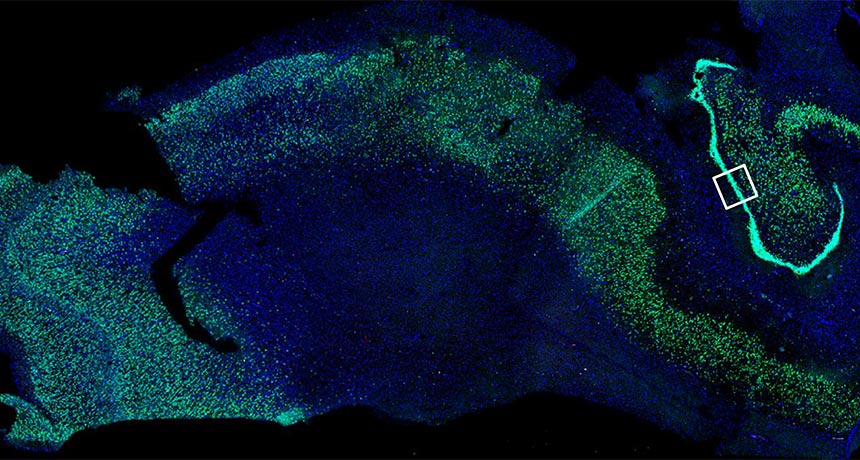
How Smilodon’s sense of smell compared with living cats’ is something else researchers wonder about. To probe this part of the extinct animal’s biology, a team lead by Van Valkenburgh looked at Smilodon’s cribriform plate — a small, perforated bone inside the skull. Smell-sensing nerve cells pass through holes in the plate from the olfactory receptors in the nose to the brain. The size and number of holes are thought to correlate with the number of receptors and, therefore, the extent of an animal’s sense of smell.

To confirm this link, Van Valkenburgh’s team combined CT scans and 3-D images of skulls from 27 species of living mammals with information on the number of olfactory receptor genes. A CT scan of a skull revealed that Smilodon may have had slightly fewer olfactory receptor nerve cells than a domestic cat, the researchers reported at the paleontology conference. Smilodon’s sense of smell was perhaps 10 to 20 percent less keen than a modern lion’s, says Van Valkenburgh, whose team reported the findings in the March 14, 2018 Proceedings of the Royal Society B.

Smilodon “might have relied more heavily on their eyes and their ears,” she says. Perhaps, in an ancient evolutionary divergence, Smilodon’s level of reliance on smell went in a slightly different direction than in modern big cats.

Saber-toothed swan song
As the pieces of the Smilodon puzzle fall into place, perhaps the biggest remaining mystery is why the animal disappeared 10,000 to 12,000 years ago. Debate about the extinction of some of North America’s large mammal species swings between blaming humans and climate change (SN: 11/10/18, p. 28). While humans, who probably arrived on the continent more than 15,000 years ago, and Smilodon certainly knew one another in the Americas, they may not have overlapped at La Brea, Shaw says. The earliest evidence of people in the Los Angeles Basin is about 11,000 years ago, by which time Smilodon may or may not already have gone. Nevertheless, human hunting of large prey elsewhere in the Americas could have led to a scarcity of food for the big cats, he says.

One theory holds that Smilodon went through tough times at La Brea when lack of prey forced the saber-toothed cats to consume entire carcasses including bones. This has been posited as the reason for all those broken teeth among the La Brea fossils. But DeSantis isn’t convinced; she thinks breakages happened during scuffles with prey. She says dental microwear suggests that Smilodon was not eating great quantities of bone.
Some opportunistic carnivores, such as cougars, did eat bone and managed to survive to the modern day. Perhaps Smilodon couldn’t adapt to hunting smaller prey when larger herbivores disappeared, also around 10,000 to 12,000 years ago (SN: 11/24/18, p. 22).

“A lot of the large prey on the landscape go extinct,” DeSantis says. “You lose out on the horses, camels, giant ground sloths, mammoths and mastodon. That’s got to have had an impact.”

The challenge of dating fossils from the tar pits has been one hurdle to understanding exactly what was going on with Smilodon over time. Bones deposited over many thousands of years get jumbled by movement in the tar, for reasons experts don’t fully understand. Plus, the tar itself becomes embedded in each specimen, complicating carbon dating.

However, new methods of chemically pretreating fossils to remove the tar have made carbon dating much easier and cheaper — and a multi-institutional project is now dating hundreds of Smilodon and other bones. Researchers will soon be able to track changes in Smilodon over the 35,000 years of prehistory recorded at La Brea and correlate fossil changes to known changes in climate over that time.

“We’re going to have a much better handle,” Van Valkenburgh says, “on what was going on towards the end of their existence.”

This article appears in the March 30, 2019 issue of Science News with the headline, “The Baddest Cat of All: Fresh details say saber-toothed Smilodon helped injured pack members.”

Signs of new nerve cells spotted in adult brains

A tweaked laboratory protocol has revealed signs of thousands of newborn nerve cells in the brains of adults, including an octogenarian.

These immature neurons, described online March 25 in Nature Medicine, mark the latest data points in the decades-old debate over whether people’s brains churn out new nerve cells into adulthood. The process, called neurogenesis, happens in the brains of some animals, but scientists have been divided over whether adult human brains are capable of such renewal (SN Online: 12/20/18).
Researchers viewed slices of postmortem brains of 13 formerly healthy people aged 43 to 87 under a microscope, and saw thousands of what appeared to be newborn nerve cells. These cells were in a part of the hippocampus called the dentate gyrus, a suspected hot spot for new neurons. Brain samples from 45 people with Alzheimer’s disease, however, had fewer of these cells — a finding that suggests that neurogenesis might also be related to the neurodegenerative disease.

Most of the brain samples used in the study were processed within 10 hours of a donor’s death, and spent no more than 24 hours soaking in a chemical that preserves the tissue. Those factors may help explain why the new neurons were spotted, the researchers write. Some earlier experiments that didn’t find evidence of neurogenesis used samples that were processed later after a donor’s death, and that had sat for longer in the fixing chemical.

Here are 5 RNAs that are stepping out of DNA’s shadow

DNA is the glamour molecule of the genetics world. Its instructions are credited with defining appearance, personality and health. And the proteins that result from DNA’s directives get credit for doing most of the work in our cells. RNA, if mentioned at all, is considered a mere messenger, a go-between — easy to ignore. Until now.

RNAs, composed of strings of genetic letters called nucleotides, are best known for ferrying instructions from the genes in our DNA to ribosomes, the machines in cells that build proteins. But in the last decade or so, researchers have realized just how much more RNAs can do — how much they control, even. In particular, scientists are finding RNAs that influence health and disease yet have nothing to do with being messengers.

The sheer number and variety of noncoding RNAs, those that don’t ferry protein-building instructions, give some clues to their importance. So far, researchers have cataloged more than 25,000 genes with instructions for noncoding RNAs in the human genome, or genetic instruction book (SN: 10/13/18, p. 5). That’s more than the estimated 21,000 or so genes that code for proteins.

Those protein-coding genes make up less than 2 percent of the DNA in the human genome. Most of the rest of the genome is copied into noncoding RNAs, and the vast majority of those haven’t been characterized yet, says Pier Paolo Pandolfi of Boston’s Beth Israel Deaconess Medical Center. “We can’t keep studying just two volumes of the book of life. We really need to study them all.”
Scientists no longer see the RNAs that aren’t envoys between DNA and ribosomes as worthless junk. “I believe there are hundreds, if not thousands, of noncoding RNAs that have a function,” says Harvard University molecular biologist Jeannie Lee. She and other scientists are beginning to learn what these formerly ignored molecules do. It turns out that they are involved in every step of gene activity, from turning genes on and off to tweaking final protein products. Those revelations were unthinkable 20 years ago.

Back in the 1990s, Lee says, scientists thought only proteins could turn genes on and off. Finding that RNAs were in charge “was a very odd concept.”

Here are five examples among the many noncoding RNAs that are now recognized as movers and shakers in the human body, for good and ill.
Sometimes anticancer drugs stop working for reasons researchers don’t entirely understand. Take the chemotherapy drug cytarabine. It’s often the first drug doctors prescribe to patients with a blood cancer called acute myeloid leukemia. But cytarabine eventually stops working for about 30 to 50 percent of AML patients, and their cancer comes back.

Researchers have looked for defects in proteins that may be the reason cytarabine and other drugs fail, but there still isn’t a complete understanding of the problem, Pandolfi says. He and colleagues now have evidence that drug resistance may stem from problems in some of the largest and most bountiful of the newly discovered classes of RNAs, known as long noncoding RNAs. Researchers have already cataloged more than 18,000 of these “lncRNAs” (pronounced “link RNAs”).
Pandolfi and colleagues investigated how some lncRNAs may work against cancer patients who are counting on chemotherapy to fight their disease. “We found hundreds of new players that can regulate response to therapy,” he says.

When the researchers boosted production of several lncRNAs in leukemia cells, the cells became resistant to cytarabine, Pandolfi and colleagues reported in April 2018 in Cell. They also found that patients with AML who had higher than normal levels of two lncRNAs experienced a cancer recurrence sooner than people who had lower levels of those lncRNAs.

Researchers are just beginning to understand how these lncRNAs influence cancer and other diseases, but Pandolfi is hopeful that someday he and other researchers will devise ways to control the bad actors and boost the helpful ones.

MicroRNAs
Sparking a tumor’s spread

MicroRNAs are barely more than 20 RNA units, or bases, long, but they play an outsized role in heart disease, arthritis and many other ailments. These pipsqueaks can also lead to nerve pain and itchiness, researchers reported last year in Science Translational Medicine and in Neuron (SN Online: 8/13/18).

Hundreds of clinical studies are testing people’s blood and tissues to determine if microRNAs can be used to help doctors better diagnose or understand conditions ranging from asthma and Alzheimer’s disease to schizophrenia and traumatic brain injury. Some researchers are beginning to develop microRNAs as drugs and seeking ways to inhibit rogue microRNAs.

So far, the little molecules’ most firmly established roles are as promoters of and protectors against cancer (SN: 8/28/10, p. 18). Pancreatic cancer, for example, is a deadly foe. Only 8.5 percent of people are still alive five years after being diagnosed with this disease, according to U.S. National Cancer Institute statistics.

Cancer biologist Brian Lewis of the University of Massachusetts Medical School in Worcester and colleagues have learned that some microRNAs spur this lethal cancer’s initial attack and help the tumor spread from the pancreas to other organs.

MicroRNAs are mirror images of portions of the messenger RNAs that shuttle protein-making instructions from DNA to the ribosomes, where proteins are built. The microRNAs pair up with their larger messenger RNA mates and slate the bigger molecules for destruction, or at least prevent their instructions from being translated into proteins. One microRNA might have hundreds of mates, or targets, through which it influences many different body functions.

Lewis studies one gang of six microRNAs, known as the miR-17~92 cluster, the first group of microRNAs found to play a role in cancer. The six normally help strike a balance between cell growth and death, but an imbalance of these little molecules can push cells toward cancer.

Tumors in pancreatic cancer patients tend to have elevated levels of the cluster. To learn what the microRNAs were doing to goad cancer into taking hold, Lewis and colleagues used a genetic trick to remove the microRNAs from the pancreas in mice that were genetically engineered to develop pancreatic tumors. Early in their lives, mice with and without the microRNA cluster had about the same number of precancerous cells.
But by the time the animals were 9 months old, a clear difference emerged. In mice with the miR-17~92 microRNAs, nearly 60 percent of the pancreas was tending toward cancer, compared with less than 20 percent in mice lacking the cluster. The finding, reported in 2017 in Oncotarget, suggests that the microRNAs aid the cancer’s start.

The researchers developed bits of RNA that block some of the cluster members from spurring on the tumor. Using human pancreatic cancer cells grown in lab dishes, Lewis and colleagues found that taking out two of the six cluster members, miR-19a and miR-19b, stopped cancer cells from forming structures called invadopodia. As their name suggests, invadopodia allow tumors to break through blood vessel walls and other barriers to spread through the body.

Transfer RNA fragments
The virus helpers

For some young children and older adults, an infection with respiratory syncytial virus, or RSV, feels like a simple cold. But each year in the United States, more than 57,000 children younger than age 5 and about 177,000 people older than 65 are hospitalized because of the virus, the U.S. Centers for Disease Control and Prevention estimates. The infection kills hundreds of babies and about 14,000 adults over 65 annually.

Slightly higher than normal levels of some microRNAs had been linked to severe RSV infections. But molecular virologist Xiaoyong Bao of the University of Texas Medical Branch in Galveston wasn’t convinced that modestly increasing amounts of a few microRNAs could really mean the difference between a child getting a slight cold and dying from the respiratory virus.

She consulted her Texas Medical Branch colleague, cancer researcher Yong Sun Lee, for advice on studying microRNAs. Lee said Bao would need to deeply examine, or sequence, RNA in cells infected with the virus. That was an expensive proposition in 2012 when Bao started the project. “But I squeezed from my [lab’s] dry bank account,” she says, to pay for the experiment. The investment paid off. Cells infected with RSV had more of one particular RNA than did uninfected cells. Surprisingly, it was a piece of a transfer RNA. Transfer RNAs, or tRNAs, are the assembly line workers of protein building. tRNAs read instructions in a messenger RNA and deliver the amino acids the ribosome needs to make a protein.
Scientists knew that working tRNAs are essential employees. Fragments, when they were found, were considered leftover bits of decommissioned tRNAs. But the fragments that Bao and colleagues discovered aren’t just worn out bits of tRNAs. Each fragment, about 30 bases long, is precisely cut from a tRNA when RSV infects cells. The fragments aid the virus’s infection in more than one way. For instance, two fragments help the virus make copies of itself in cells, Bao and colleagues reported in 2017 in the Journal of General Virology.

tRNA fragments may also boost the body’s susceptibility to a virus. Last year, Bao’s group described in Scientific Reports how exposure to some heavy metals, via air or water pollution, can produce tRNA fragments that trigger inflammation, which may make people more susceptible to respiratory infections such as RSV.

SINE RNAs
Sacrificing infected cells

Another type of RNA may help protect against infection by certain viruses, including herpesvirus. Virologist Britt Glaunsinger has long marveled at the way viruses manipulate host cells by controlling RNAs in the cell. She became intrigued by transposons, mobile stretches of DNA that can jump from one location to another in the genome. Transposons make up nearly half of all the DNA in the human genome (SN: 5/27/17, p. 22). “We tend to think of [transposons] as parasites and things our own cells are constantly trying to shut down,” says Glaunsinger, a Howard Hughes Medical Institute investigator at the University of California, Berkeley. That’s because some are relics of ancient viruses. “While they may have initially been bad, some of them may actually be useful to us,” she says.

One class of transposons, called SINEs for short interspersed nuclear elements, are peppered throughout the genome. People have more than a million of one type of SINE known as Alu elements. Mice have similar SINEs, called B2s.

When active, SINE transposons make RNA copies of themselves. These SINE RNAs don’t carry instructions for building proteins and alone don’t enable the transposons to jump around the genome. So researchers puzzled over their role. Glaunsinger and colleagues discovered that some SINE RNAs may protect against viral infections.
Normally, cells keep a tight lock on transposons, preventing them from making any RNA. But in Glaunsinger’s experiments, cells infected with herpesvirus “were producing tons of these noncoding RNAs in response to infection,” she says. “That sort of captured our interest.”

Details of the process are still being worked out, but Glaunsinger and others have discovered that SINE RNA production triggers a cascade of events that eventually kills infected human and mouse cells. Once the RNA production gets going, Glaunsinger says, “the cell is destined to die.” Inflammation appears to be an important step in the cell-killing chain reaction. It’s all for the greater good: Killing the infected cell may protect the rest of the organism from the infection’s spread.

But there’s a wrinkle: In mice, at least, one type of herpesvirus benefits from the flood of B2 RNAs in the cells it infects. The virus hijacks part of the inflammation chain reaction to boost its own production, Glaunsinger and colleagues reported in 2015 in PLOS Pathogens. “This is an example of the back-and-forth battle that’s always going on between virus and host,” she says. “Now the ball is back in the host’s court.”

piRNAs
Shielding the brain from jumping genes

Autopsies of people who died with Alzheimer’s disease show a buildup of a protein called tau in the brain. That tau accumulation is tied to loss of some guardian RNAs, according to work by Bess Frost, a neurobiologist at UT Health San Antonio.

Frost studies fruit flies genetically engineered to make a disease-causing version of human tau in their nerve cells. Flies with the disorderly tau get a progressive nerve disease that causes movement problems and kills nerves. The insects live shorter lives than normal.

Part of the reason the flies, as well as people with tau tangles, have problems is because some RNAs known to guard the genome fall down on the job, Frost and colleagues discovered. These piwi-interacting RNAs, or piRNAs (pronounced “pie RNAs”), help keep transposons from jumping around. When transposons jump, they may land in or near a gene and mess with its activity.
Usually cells prevent jumping by stopping transposons from making messenger RNA, which carries instructions to make proteins that eventually enable the transposon to hop from place to place. If a transposon gets past the cell’s defenses and produces its messenger RNA, piRNAs will step up to pair with the messenger and cause its destruction.

When disease-causing tau builds up in flies (and maybe in people), a class of transposon with a lengthy name — class I long terminal repeat retrotransposons — makes much more RNA than usual. And when flies have the disease-causing version of tau, they also have lower than normal levels of piRNAs, Frost and colleagues reported in August 2018 in Nature Neuroscience. “Both arms of control are messed up,” Frost says. Brains of people who died with Alzheimer’s disease or supranuclear palsy, another tau-related disease, also show signs that transposons were making extra RNA, suggesting that when tau goes bad, it can beat piRNA’s defenses.

In search of a work-around, Frost’s team found that genetically boosting piRNA production in flies or giving a drug that stops transposon hops reduced nerve cell death in the insects. The researchers are preparing to test the drug in mice prone to a rodent version of Alzheimer’s disease. The team is also examining human brain tissue to see if the increase in transposon RNAs actually leads to transposon jumping in Alzheimer’s patients. If transposons don’t hop more than usual, the finding may suggest that transposon RNAs themselves can cause mischief — no jumping necessary.

This story appears in the April 13, 2019 issue of Science News with the headline, “The Secret Powers of RNA: Overlooked molecules play a big role in human health.”

Mathematicians may have found the fastest way to multiply huge numbers

Multiplying 2 x 2 is easy. But multiplying two numbers with more than a billion digits each — that takes some serious computation.

The multiplication technique taught in grade school may be simple, but for really big numbers, it’s too slow to be useful. Now, two mathematicians say that they’ve found the fastest way yet to multiply extremely large figures.

The duo claim to have achieved an ultimate speed limit for multiplication, first suggested nearly 50 years ago. That feat, described online March 18 at the document archive HAL, has not yet passed the gauntlet of peer review. But if the technique holds up to scrutiny, it could prove to be the fastest possible way of multiplying whole numbers, or integers.
If you ask an average person what mathematicians do, “they say, ‘Oh, they sit in their office multiplying big numbers together,’” jokes study coauthor David Harvey of the University of New South Wales in Sydney. “For me, it’s actually true.”

When making calculations with exorbitantly large numbers, the most important measure of speed is how quickly the number of operations needed — and hence the time required to do the calculation — grows as you multiply longer and longer strings of digits.

That growth is expressed in terms of n, defined as the number of digits in the numbers being multiplied. For the new technique, the number of operations required is proportional to n times the logarithm of n, expressed as O(n log n) in mathematical lingo. That means that, if you double the number of digits, the number of operations required will increase a bit faster, more than doubling the time the calculation takes.
But, unlike simpler methods of multiplication, the time needed doesn’t quadruple, or otherwise rapidly blow up, as the number of digits creeps up, report Harvey and Joris van der Hoeven of the French national research agency CNRS and École Polytechnique in Palaiseau. That slower growth rate makes products of bigger numbers more manageable to calculate.

The previously predicted max speed for multiplication was O(n log n), meaning the new result meets that expected limit. Although it’s possible an even speedier technique might one day be found, most mathematicians think this is as fast as multiplication can get.

“I was very much astonished that it had been done,” says theoretical computer scientist Martin Fürer of Penn State. He discovered another multiplication speedup in 2007, but gave up on making further improvements. “It seemed quite hopeless to me.”

The new technique comes with a caveat: It won’t be faster than competing methods unless you’re multiplying outrageously huge numbers. But it’s unclear exactly how big those numbers have to be for the technique to win out — or if it’s even possible to multiply such big numbers in the real world.

In the new study, the researchers considered only numbers with more than roughly 10214857091104455251940635045059417341952 digits when written in binary, in which numbers are encoded with a sequence of 0s and 1s. But the scientists didn’t actually perform any of these massive multiplications, because that’s vastly more digits than the number of atoms in the universe. That means there’s no way to do calculations like that on a computer, because there aren’t enough atoms to even represent such huge numbers, much less multiply them together. Instead, the mathematicians came up with a technique that they could prove theoretically would be speedier than other methods, at least for these large quantities.

There’s still a possibility that the method could be shown to work for smaller, but still large, numbers. That could possibly lead to practical uses, Fürer says. Multiplication of these colossal numbers is useful for certain detailed calculations, such as finding new prime numbers with millions of digits (SN Online: 1/5/18) or calculating pi to extreme precision (SN Online: 12/10/02).

Even if the method is not widely useful, making headway on a problem as fundamental as multiplication is still a mighty achievement. “Multiplying numbers is something people have been working on for a while,” says mathematical physicist John Baez of the University of California, Riverside. “It’s a big deal, just because of that.”

Plant/animal hybrid proteins could help crops fend off diseases

A new biological mashup just dropped.

“Pikobodies,” bioengineered immune system proteins that are part plant and part animal, could help flora better fend off diseases, researchers report in the March 3 Science. The protein hybrids exploit animals’ uniquely flexible immune systems, loaning plants the ability to fight off emerging pathogens.

Flora typically rely on physical barriers to keep disease-causing microbes at bay. If something unusual makes it inside the plants, internal sensors sound the alarm and infected cells die. But as pathogens evolve ways to dodge these defenses, plants can’t adapt in real time. Animals’ adaptive immune systems can, making a wealth of antibodies in a matter of weeks when exposed to a pathogen.、
In a proof-of-concept study, scientists genetically modified one plant’s internal sensor to sport animal antibodies. The approach harnesses the adaptive immune system’s power to make almost unlimited adjustments to target invaders and lends it to plants, says plant immunologist Xinnian Dong, a Howard Hughes Medical Institute investigator at Duke University who was not involved in the work.

Crops especially could benefit from having more adaptable immune systems, since many farms grow fields full of just one type of plant, says Dong. In nature, diversity can help protect vulnerable plants from disease-spreading pathogens and pests. A farm is more like a buffet.

Researchers have had success fine-tuning plant genes to be disease-resistant, but finding the right genes and editing them can take more than a decade, says plant pathologist Sophien Kamoun of the Sainsbury Laboratory in Norwich, England. He and colleagues wanted to know if plant protection could get an additional boost from animal-inspired solutions.

To create the pikobodies, the team fused small antibodies from llamas and alpacas with a protein called Pik-1 that’s found on the cells of Nicotiana benthamiana, a close relative of tobacco plants. Pik-1 typically detects a protein that helps a deadly blast fungus infect plants (SN: 7/10/17). For this test, the animal antibodies had been engineered to target fluorescent proteins

Plants with the pikobodies killed cells exposed to the fluorescent proteins, resulting in dead patches on leaves, the team found. Of 11 tested versions, four were not toxic to the leaves and triggered cell death only when the pikobodies attached to the specific protein that they had been designed bind.

What’s more, pikobodies can be combined to give plants more than one way to attack a foreign invader. That tactic could be useful to hit pathogens with the nimble ability to dodge some immune responses from multiple angles.

Theoretically, it’s possible to make pikobodies “against virtually any pathogen we study,” Kamoun says. But not all pikobody combos worked together in tests. “It’s a bit hit or miss,” he says. “We need some more basic knowledge to improve the bioengineering.”

Southern right whale moms and calves may whisper to evade orcas

Whales are known for belting out sounds in the deep. But they may also whisper.

Southern right whale moms steer their calves to shallow waters, where newborns are less likely to be picked off by an orca. There, crashing waves mask the occasional quiet calls that the pairs make. That may help the whales stick together without broadcasting their location to predators, researchers report July 11 in the Journal of Experimental Biology.

While most whale calls are meant to be long-range, “this shows us that whales have a sort of intimate communication as well,” says Mia Nielsen, a behavioral biologist at Aarhus University in Denmark. “It’s only meant for the whale right next to you.”

Nielsen and colleagues tagged nine momma whales with audio recorders and sensors to measure motion and water pressure, and also recorded ambient noise in the nearshore environment. When the whales were submerged, below the noisy waves, the scientists could pick up the hushed calls, soft enough to fade into the background noise roughly 200 meters away.
An orca, or killer whale, “would have to get quite close in the big ocean to be able to detect them,” says biologist Peter Tyack at the University of St. Andrews in Scotland. Tyack was not involved with the study, but collaborates with one of the coauthors on other projects.

The whispers were associated with times when the whales were moving, rather than when mothers were stationary and possibly suckling their calves. Using hushed tones could make it harder for the pair to reunite if separated. But the observed whales tended to stay close to one another, about one body length apart, the team found.

Eavesdropping biologists have generally focused on the loud noises animals make, Tyack says. “There may be a repertoire among the calls of lots of animals that are specifically designed only to be audible to a partner who’s close by,” he says.

The standard model of particle physics passed one of its strictest tests yet

No one has ever probed a particle more stringently than this.

In a new experiment, scientists measured a magnetic property of the electron more carefully than ever before, making the most precise measurement of any property of an elementary particle, ever. Known as the electron magnetic moment, it’s a measure of the strength of the magnetic field carried by the particle.

That property is predicted by the standard model of particle physics, the theory that describes particles and forces on a subatomic level. In fact, it’s the most precise prediction made by that theory.
By comparing the new ultraprecise measurement and the prediction, scientists gave the theory one of its strictest tests yet. The new measurement agrees with the standard model’s prediction to about 1 part in a trillion, or 0.1 billionths of a percent, physicists report in the February 17 Physical Review Letters.

When a theory makes a prediction at high precision, it’s like a physicist’s Bat Signal, calling out for researchers to test it. “It’s irresistible to some of us,” says physicist Gerald Gabrielse of Northwestern University in Evanston, Ill.

To measure the magnetic moment, Gabrielse and colleagues studied a single electron for months on end, trapping it in a magnetic field and observing how it responded when tweaked with microwaves. The team determined the electron magnetic moment to 0.13 parts per trillion, or 0.000000000013 percent.

A measurement that exacting is a complicated task. “It’s so challenging that nobody except the Gabrielse team dares to do it,” says physicist Holger Müller of the University of California, Berkeley.
The new result is more than twice as precise as the previous measurement, which stood for over 14 years, and which was also made by Gabrielse’s team. Now the researchers have finally outdone themselves. “When I saw the [paper] I said, ‘Wow, they did it,’” says Stefano Laporta, a theoretical physicist affiliated with University of Padua in Italy, who works on calculating the electron magnetic moment according to the standard model.

The new test of the standard model would be even more impressive if it weren’t for a conundrum in another painstaking measurement. Two recent experiments, one led by physicist Saïda Guellati-Khélifa of Kastler Brossel Laboratory in Paris and the other by Müller, disagree on the value of a number called the fine-structure constant, which characterizes the strength of electromagnetic interactions (SN: 4/12/18). That number is an input to the standard model’s prediction of the electron magnetic moment. So the disagreement limits the new test’s precision. If that discrepancy were sorted out, the test would become 10 times as precise as it is now.
The stalwart standard model has stood up to a barrage of experimental tests for decades. But scientists don’t think it’s the be-all and end-all. That’s in part because it doesn’t explain observations such as the existence of dark matter, an invisible substance that exerts gravitational influence on the cosmos. And it doesn’t say why the universe contains more matter than antimatter (SN: 9/22/22). So physicists keep looking for cases where the standard model breaks down.

One of the most tantalizing hints of a failure of the standard model is the magnetic moment not of the electron, but of the muon, a heavy relative of the electron. In 2021, a measurement of this property hinted at a possible mismatch with standard model predictions (SN: 4/7/21).

“Some people believe that this discrepancy could be the signature of new physics beyond the standard model,” says Guellati-Khélifa, who wrote a commentary on the new electron magnetic moment paper in Physics magazine. If so, any new physics affecting the muon could also affect the electron. So future measurements of the electron magnetic moment might also deviate from the prediction, finally revealing the standard model’s flaws.

Young black holes evade detection

HONOLULU — Perhaps most supermassive black holes — dark giants in the centers of galaxies — are just shy when they’re young.

“We have this weird problem, where on the one hand the universe makes really supermassive black holes very shortly after the Big Bang,” says Kevin Schawinski, an astrophysicist at ETH Zürich in Switzerland. “But when we look at more typical galaxies, we find no evidence for growing black holes.”

The feeding zones around voracious black holes create quasars, blazing furnaces of X-rays and other light. And yet the Chandra space telescope detects no X-rays from a cache of galaxies in the constellation Fornax that researchers think should be nourishing young black holes, Schawinski reported August 6 at a meeting of the International Astronomical Union.
Over the past several years, astronomers have found a handful of very bright quasars that lit up within the first billion years of cosmic history. These quasars are probably powered by unusually hefty supermassive black holes — ones that gobbled down gas as fast as physically possible (or even faster) for hundreds of millions of years.

“If this happens all over the universe,” says Schawinski, “then if we look at more normal-mass galaxies, we should be seeing their supermassive black holes pop out in the early universe to the same degree.”

But they don’t.
Maybe the more run-of-the-mill black holes are there but they’re not actively feeding, he says. Or perhaps something is blocking the X-rays from getting out.

Or maybe — just maybe — these black holes haven’t been born yet.

“It’s a very interesting suggestion,” says Andrea Comastri, an astronomer at the Osservatorio Astronomico di Bologna in Italy, says of the not-yet-born scenario. “But I’m not convinced.”

These images capture a relatively tiny volume of space, he says, so perhaps the researchers aren’t casting a wide enough net. The distances to these galaxies are also notoriously difficult to pin down. Many could be much closer and seen during a time when black holes have formed but quieted down a bit.

If the universe can make monstrous black holes in under a billion years, then making the relatively little guys should be straightforward and they should be everywhere, Comastri says. “It should be easier to make smaller black holes because you don’t have to work that much. They are there somewhere.”

If the black holes are confirmed to be missing, “it’s going to shake up a lot of what we think about the growth of quasars,” says Tiziana Di Matteo, an astrophysicist at Carnegie Mellon University in Pittsburgh. “But I’m very skeptical of it.”

These cosmic no-shows probably don’t suck down gas as fast as the researchers assume, she says. If these black holes only nibble at the surrounding gas — as opposed to their obese cousins who gorge themselves — then X-rays would only trickle from their dinner plates and might not be detected.

Much like with humans, black hole obesity is influenced by environment. Most galaxies need some time to build up enough mass to efficiently feed their black holes, Di Matteo says. Tiny galaxies easily lose gas every time a cluster of new stars is born or whenever a dying star explodes. “It’s only in extreme environments,” she says, at the junctions of cosmic filaments that become interstellar dumping grounds, “where gas could plunge through and not care about anything else that’s going on.” Here, fledgling black holes aren’t as reliant on their galaxy’s feeble gravity to grab food; the incoming rivers of gas are like intergalactic fire hoses.

Those unusually massive black hole starter kits are probably responsible for the dazzling quasars that switch on during the first billion years after the Big Bang. Computer simulations show that in the younger, more intimate universe, when everything was squished together a lot more than today, there are the oddball places where gas funnels onto ancestral galaxies at astounding rates, providing fast-growing black holes with an all-you-can-eat buffet.

The other less showy black holes, the ones Schawinski and colleagues are hunting for, probably spend the next several billion years quietly catching up. Finding these black holes when they’re young and struggling to grow might require searching a wider area or getting more sensitive observations.

“It’s exciting,” Schawinski says. “It’s the last major category of astrophysical objects of whose origin we know nothing about.” Planets, stars and galaxies are pretty well understood, he says. “But we have no idea how supermassive black holes form.”

Schawinski’s team plans to spend the next year or two repeating their experiment over a wider volume of space, hoping to find at least one youthful black hole in a moderate-sized galaxy. “Once you go from zero to one you have something to work with,” he says. “Right now we’ve got nothing.”

Chemical residue reveals ancient Egyptians’ mummy-making mixtures

Scientists have unwrapped long-sought details of embalming practices that ancient Egyptians used to preserve dead bodies.

Clues came from analyses of chemical residue inside vessels from the only known Egyptian embalming workshop and nearby burial chambers. Mummification specialists who worked there concocted specific mixtures to embalm the head, wash the body, treat the liver and stomach, and prepare bandages that swathed the body, researchers report February 1 in Nature.

“Ancient Egyptian embalmers had extensive chemical knowledge and knew what substances to put on the skin to preserve it, even without knowing about bacteria and other microorganisms,” Philipp Stockhammer, an archaeologist at Ludwig Maximilians University of Munich, said at a January 31 news conference.
The findings come courtesy of chemical residue inside 31 vessels found in an Egyptian embalming workshop and four vessels discovered in an adjacent pair of burial chambers. Writing on workshop vessels named embalming substances, provided embalming instructions (such as “to put on his head”) or both. All the artifacts — dating from Egypt’s 26th dynasty which rose to power between 664 B.C. and 525 B.C. — were excavated at a cemetery site called Saqqara in 2016. Archaeologist and study coauthor Ramadan Hussein, who died in 2022, led that project.

Newfound mummy embalming mixtures
Five of the vessels had the label antiu. The substance was thought to have been a fragrant resin called myrrh. The antiu at Saqqara, however, consisted of oil or tar from cedar and juniper or cypress trees mixed with animal fats. Writing on these jars indicates that antiu could have been used alone or combined with another substance called sefet.

Three vessels from the embalming workshop bore the label sefet, which researchers have usually described as an unidentified oil. At Saqqara, sefet was a scented, fat-based ointment with added ingredients from plants. Two sefet pots contained animal fats mixed with oil or tar from juniper or cypress trees. A third container held animal fats and elemi, a fragrant resin from tropical trees.

Clarification of the ingredients in antiu and sefet at Saqqara “takes mummification studies further than before,” says Egyptologist Bob Brier of Long Island University in Brookville, N.Y., who was not part of the research.

Egyptians may have started mummifying their dead as early as 6,330 years ago (SN: 8/18/14). Mummification procedures and rituals focused on keeping the body fresh so the deceased could enter what was believed to be an eternal afterlife.
Embalming and mummification procedures likely changed over time, says team member Maxime Rageot, a biomolecular archaeologist also at Ludwig Maximilians University. Embalmers’ mixtures at Saqqara may not correspond, say, to those used around 700 years earlier for King Tutankhamun (SN: 11/2/22).

Mummy embalming instructions
Outside surfaces of other vessels from the Saqqara embalming workshop and burial chambers sported labels and, in some cases, instructions for treatment of the head, preparation of linen mummy bandages, washing the body and treating the liver and stomach. Inscriptions on one jar referred to an administrator who performed embalming procedures, mainly on the head.

Chemical residue inside these pots consisted of mixtures specific to each embalming procedure. Ingredients included oils or tars of cedar and juniper or cypress trees, pistachio resin, castor oil, animal fats, heated beeswax, bitumen (a dense, oily substance), elemi and a resin called dammar.

Most of those substances have been identified in earlier studies of chemical residues from Egyptian mummies and embalming vessels in individual tombs, says Egyptologist Margaret Serpico of University College London. But elemi and dammar resins have not previously been linked to ancient Egyptian embalming practices and are “highly unexpected,” notes Serpico, who did not participate in the new study.

Elemi was an ingredient in the workshop mixtures used to treat the head, the liver and bandages wrapped around the body. Chemical signs of dammar appeared in a vessel from one of the burial chambers that included remnants of a range of substances, indicating that the container had been used to blend several different mixtures, the researchers say.

Specific properties of elemi and dammar that aided in preserving dead bodies have yet to be investigated, Stockhammer said.

A far-flung trade network for mummy embalming ingredients
Elemi resin reached Egypt from tropical parts of Africa or Southeast Asia, while dammar originated in Southeast Asia or Indonesia, Rageot says. Other embalming substances detected at Saqqara came from Southwest Asia and parts of southern Europe and northern Africa bordering the Mediterranean Sea. These findings provide the first evidence that ancient Egyptian embalmers depended on substances transported across vast trade networks.

Egyptian embalmers at Saqqara took advantage of a trade network that already connected Egypt to sites in Southeast Asia, Stockhammer said. Other Mediterranean and Asian societies also engaged in long-distance trade during ancient Egypt’s heyday (SN: 1/9/23).

It’s no surprise that ancient Egyptians imported embalming ingredients from distant lands, Brier says. “They were great traders, had limited [local] wood products and really wanted these substances to achieve immortality.”

Some young sea spiders can regrow their rear ends

No backside, no problem for some young sea spiders.

The creatures can regenerate nearly complete parts of their bottom halves — including muscles, reproductive organs and the anus — or make do without them, researchers report January 23 in Proceedings of the National Academy of Sciences.

The ability to regrow body parts isn’t super common, but some species manage to pull it off. Some sea slug heads can craft an entirely new body (SN: 3/8/21). Sea spiders and some other arthropods — a group of invertebrates with an exoskeleton — can regrow parts of their legs. But researchers thought new legs were the extent of any arthropod’s powers, perhaps because tough exteriors somehow stop them from regenerating other body parts.
A mishap first clued evolutionary biologist Georg Brenneis in that sea spiders (Pycnogonum litorale) might be able handle more complex repairs too. He accidentally injured one young specimen that he was working on in the lab with forceps. “It wasn’t dead, it was moving, so I just kept it,” says Brenneis, of the University of Vienna. Several months later, the sea spider had an extra leg instead of a scar, he and evolutionary biologist Gerhard Scholtz of Humbolt University of Berlin reported in 2016 in The Science of Nature.

In the new study, most of the 19 young spiders recovered and regrew missing muscles and other parts of their lower halves after amputation, though the regeneration wasn’t always perfect. Some juveniles sported six or seven legs instead of eight.

None of four adults regenerated. That may be because adults no longer shed their skin as they grow, suggesting that regeneration and molting are somehow linked, Brenneis says. Two young sea spiders also didn’t regenerate at all. The animals survived with only four legs and without an anus. Instead of pooping, the pair regurgitated waste out of their mouths.
Next up is figuring out whether other arthropods also regenerate more than scientists thought, and how sea spiders do it, Brenneis says. “I would like to see how it works.”