Riding roller coasters might help dislodge kidney stones
Passing a kidney stone is not exactly rocket science, but it could get a boost from Space Mountain.
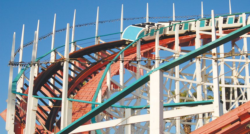
It seems that shaking, twisting and diving from on high could help small stones dislodge themselves from the kidney’s inner maze of tubules. Or so say two researchers who rode the Big Thunder Mountain Railroad roller coaster at Disney’s Magic Kingdom in Orlando, Fla., 20 times with a fake kidney tucked inside a backpack.
The researchers, from Michigan State University College of Osteopathic Medicine in East Lansing, planned the study after several of their patients returned from the theme park announcing they had passed a kidney stone. Finally, one patient reported passing three stones, each one after a ride on a roller coaster.
“Three consecutive rides, three stones — that was too much to ignore,” says David Wartinger, a kidney specialist who conducted the study with Marc Mitchell, his chief resident at the time.
Since neither of the two had kidney stones themselves, the pair 3-D printed a life-size plastic replica of the branching interior of a human kidney. Then they inserted three stones and human urine into the model. The stones were of the size that usually pass on their own, generally smaller in diameter than a grain of rice. After arriving at the park, Wartinger and Mitchell sought permission from guest services to do the research, fearing that two men with a backpack boarding the same ride over and over might strike workers as suspect.
“Luckily, the first person we talked to in an official capacity had just passed a kidney stone,” Wartinger says. “He told us he would help however we needed.”
Even when a stone is small, its journey through the urinary tract can be excruciating. In the United States alone, more than 1.6 million people each year experience kidney stones painful enough to send them to the emergency room. Larger stones — say, the size of a Tic Tac — can be treated with sound waves that break the stones into smaller pieces that can pass.
For the backpack kidney, the rear of the train was the place to be. About 64 percent of the stones in the model kidney cleared out after a spin in the rear car. Only about 17 percent passed after a single ride in the front car, the researchers report in the October Journal of the American Osteopathic Association.
Wartinger thinks that a coaster with more vibration and less heart-pounding speed would be better at coaxing a stone on its way.
The preliminary study doesn’t show whether real kidneys would yield their stones to Disney magic. Wartinger says a human study would be easy and inexpensive, but for now, it’s probably wise to check with a doctor before taking the plunge.