Young penguins follow false food cues

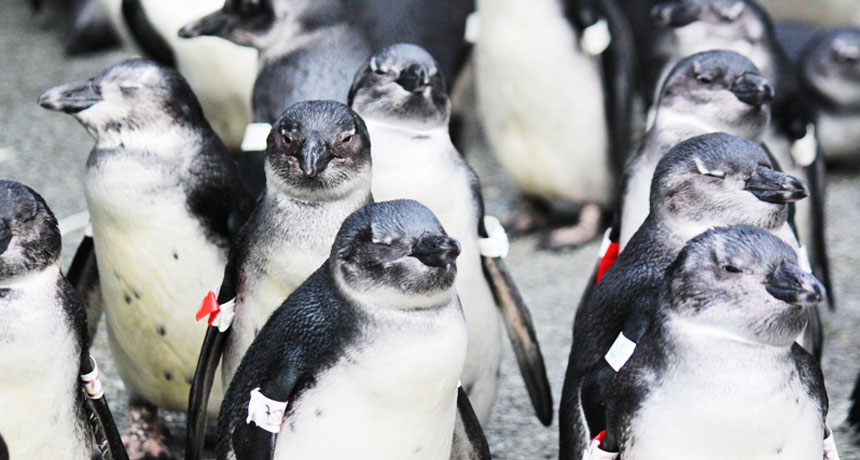
African penguins have used biological cues in the ocean for centuries to find their favorite fish. Now these cues are trapping juvenile penguins in areas with hardly any food, scientists report February 9 in Current Biology.

It’s the first known ocean “ecological trap,” which occurs when a once-reliable environmental cue instead, often because of human interference, prompts an animal to do something harmful.

When juvenile Spheniscus demersus penguins off the Western Cape of South Africa leave the nest for their maiden voyage at sea, they head for prime penguin hunting grounds. But the fish are no longer there, says Richard Sherley, a marine ecologist with the University of Exeter Environment and Sustainability Institute. Increased ocean temperatures, changes in salinity and overfishing have driven the fish eastward.
Penguins are doing what they’ve evolved to do, following signs in the water to historically prosperous habitats. “But humans have broken the system,” Sherley says, and there’s no longer enough fish to support the seabirds.

Sherley estimates that only about 20 percent of these African penguins survive their first year, partly because they can fall into this ecological trap.
Ecological traps have been documented on land for decades. There has been a lot of speculation about traps in the ocean, but this study is the best evidence so far, says Rob Hale, an ecologist with the University of Melbourne.
“Hopefully the study will generate more interest in examining ecological traps in the ocean so we can better understand when and why traps arise, how they are likely to affect animals, and how we can go about managing their effects,” Hale says.

This trap may have occurred because of how penguins find their food. Researchers think penguins can sense a stress chemical that phytoplankton release when being eaten. Penguins eat sardines, which eat phytoplankton. Usually the chemical, dimethyl sulfide, signals to penguins where the fish are feasting on phytoplankton. But phytoplankton can release the compound in other situations, like in rough water. The signal is still sent, but there are no fish.

“You have a cue that used to signal high quality in an environment, but that environment has been modified by human action to some extent,” Sherley says. “The animals are tricked or trapped into selecting a lower quality habitat because the cue still exists, even though there’s high quality habitat available.”

Adult penguins have adapted to the trap and shifted their hunting patterns to follow the fish east. Sherley says they’re not sure how adults learned to avoid the problem, but that there must be a way that juveniles who survive to adulthood also adapt.

Researchers also tracked juvenile penguins from the Namibia and Eastern Cape of South Africa breeding regions. The eastern penguins have been unaffected by the trap because the fish have moved closer to them. The Namibia population is being barely sustained by the goby, a junk food fish that appears to be taking over the areas previously inhabited by sardines and anchovies.

The Western Cape penguins have been most affected. The population has declined 80 percent in the last 15 years — from 40,000 breeding pairs to 5,000 or 6,000, Sherley says. He estimates that if juvenile penguins hadn’t been falling victim to this trap, the Western Cape population would be double its current levels.

If the loss of fish off the Western Cape of South Africa can’t be reversed, Sherley speculates the two most likely outcomes are an African penguin extinction or an ecosystem shift. Current penguin conservation efforts protect penguin breeding areas, but the study suggests that the protections may be insufficient because the ecological trap is far from the breeding grounds.

Europa lander mission takes another step toward reality

BOSTON — For the first time since the Viking missions to Mars in the 1970s, NASA is making the search for evidence of life on another world the primary science goal of a space mission. The target world is Jupiter’s moon Europa, considered possibly habitable because of its subsurface ocean.

The proposed mission, which could be operational in the next two decades, calls for a lander with room for roughly 43 kilograms of science instruments. They include a robotic arm to scoop samples and others to analyze the chemistry of the Jovian moon’s icy surface (SN: 5/17/14, p. 20). “It’s the first time in human history that we have the ability to design instruments to detect life within our own solar system’s backyard in the next 20 years,” astrobiologist and planetary scientist Kevin Hand said February 17 at the annual meeting of the American Association for the Advancement of Science. Hand’s team submitted a 264-page report describing the potential mission to NASA on February 8. The report is now open for review by the scientific community.

A major concern is taking precautions to prevent contamination of Europa by microbes from Earth. “These are important for protecting Europa for Europans,” said Hand, who works out of NASA’s Jet Propulsion Laboratory in Pasadena, Calif. His team proposes baking the spacecraft to kill as many microorganisms clinging to the exterior of the lander as possible.

Decontamination precautions are important not only for protecting the life that’s on the world being explored, notes biologist Norine Noonan of the University of South Florida St. Petersburg. They are also important for the science goals of the potential mission. “You don’t want to send a $2 billion spacecraft somewhere to discover E. coli,” Noonan says.

Origin of photosynthesis may go further back than estimates from 50 years ago

From deep in the gold mines of South Africa’s Orange Free State has come evidence that there was some form of biologic activity on Earth at least 2.15 billion years ago. Polymerized hydrocarbon “chemo-fossils” found in the gold ores … [probably] were originally part of a rich bacterial and algal life in the Witwatersrand basin. Since the rock layers from which they come have been dated to about 2.15 billion years ago, it seems likely that photosynthesis existed on Earth before then. — Science News, March 18, 1967

UPDATE
Scientists still debate when early photo­synthesizing organisms called cyanobacteria began pumping oxygen into Earth’s atmosphere. Recent evidence suggests the microbes existed some 3.2 billion years ago (SN Online: 9/8/15), even though a larger oxygen surge didn’t happen until about 2.4 billion years ago (SN: 3/4/17 p. 9). Those tiny bacteria left an outsized impact on our planet, releasing extra oxygen into the atmosphere that paved the way for complex multicellular life like plants and animals.

Scratching is catching in mice

Catch sight of someone scratching and out of nowhere comes an itch, too. Now, it turns out mice suffer the same strange phenomenon.

Tests with mice that watched itchy neighbors, or even just videos of scratching mice, provide the first clear evidence of contagious scratching spreading mouse-to-mouse, says neuroscientist Zhou-Feng Chen of Washington University School of Medicine in St. Louis. The quirk opens new possibilities for exploring the neuroscience behind the spread of contagious behaviors.
For the ghostly itch, experiments trace scratching to a peptide nicknamed GRP and areas of the mouse brain better known for keeping the beat of circadian rhythms, Chen and colleagues found. They report the results in the March 10 Science.

In discovering this, “there were lots of surprises,” Chen says. One was that mice, nocturnal animals that mostly sniff and whisker-brush their way through the dark, would be sensitive to the sight of another mouse scratching. Yet Chen had his own irresistible itch to test the “crazy idea,” he says.

Researchers housed mice that didn’t scratch any more than normal within sight of mice that flicked and thumped their paws frequently at itchy skin. Videos recorded instances of normal mice looking at an itch-prone mouse mid-scratch and, shortly after, scratching themselves. In comparison, mice with not-very-itchy neighbors looked at those neighbors at about the same frequency but rarely scratched immediately afterward.
Videos of scratching mice produced the same result. More audience itching and scratching followed a film of a mouse with itchy skin than one of a mouse poking about on other rodent business.
Next, researchers looked at how contagious itching plays out in the mouse nervous system. Brains of mice recently struck by contagious urges to scratch showed heightened activity in several spots, including, surprisingly, a pair of nerve cell clusters called the suprachiasmatic nuclei, or SCN. People have these clusters, too, deep in the brain roughly behind the eyes.

Other tests linked the contagious itching with GRP, previously identified as transmitting itch information elsewhere in the mouse nervous system. Mice didn’t succumb to contagious itching if they had no working genes for producing GRP or the molecule that detects it. Yet these mice still scratched when researchers irritated their skin. Also, in normal mice, a dose of GRP injected to the SCN brain regions brought on scratching without the sight of an itchy neighbor, but a dose of plain saline solution to same spots failed to set off much pawing.

It’s fine work, says dermatologist Gil Yosipovitch, who studies itching at the University of Miami. But he wonders how the mouse discovery might apply to people. So far, brain imagery in his own work has not turned up evidence for an SCN role in human contagious itching, he says.

SCN is better known as a circadian timekeeper, responding to cues in light. It’s unclear how the nerve cell clusters might orchestrate behavior based on seeing a scratching mouse, “a very specific and rich visual stimulus,” says psychologist and neuroscientist Henning Holle of the University of Hull in England. Other research suggests different brain regions are involved in contagious itching in people.

Tracking down the mechanisms behind the phenomenon is more than an intriguing science puzzle, Yosipovitch says. People troubled with strong, persistent itching are often unusually susceptible to contagious scratching, and new ideas for easing their misery would be welcome.

Life on Earth may have begun as dividing droplets

NEW ORLEANS — In a primordial soup on ancient Earth, droplets of chemicals may have paved the way for the first cells. Shape-shifting droplets split, grow and split again in new computer simulations. The result indicates that simple chemical blobs can exhibit replication, one of the most basic properties of life, physicist Rabea Seyboldt of the Max Planck Institute for the Physics of Complex Systems in Dresden, Germany, reported March 16 at a meeting of the American Physical Society.

Within a liquid, small droplets of particular chemicals can separate out, like beads of oil in water. Such globules typically remain spherical, growing as they merge with other drops. But in simulations, Seyboldt and colleagues found that droplets might behave in a counterintuitive way under certain conditions, elongating and eventually dividing into two.
If additional droplet material is continuously produced in reactions in the primordial soup, chemicals will accumulate on either end of a droplet, causing it to elongate, the simulations show. Meanwhile, waste products from the droplet are eliminated from the middle, causing the droplet to pinch in and eventually split. The resulting pair of droplets would then grow and split again to create a new generation. In addition to the above reactions, the process requires an energy source, such as heat or chemicals from a hydrothermal vent, to get reactions going.

The study, which was also described in Nature Physics in December, is theoretical — the researchers didn’t select particular chemicals for study but simply showed that certain types of reactions could cause droplets to split.

How such droplets would have evolved into vastly more complicated cells is unknown. “This is really a minimal scenario that’s supposed to give the very first indications of something that goes towards life, but if you look at living cells today, they’re infinitely more complex,” Seyboldt said.

Camera trap catches a badger burying a cow

The American badger is known to cache carrion in the ground. The animals squirrel away future meals underground, which acts something like a natural refrigerator, keeping their food cool and hidden from anything that might want to steal it. Researchers, though, had never spotted badgers burying anything bigger than a jackrabbit — until 2016, when a young, dead cow went missing in a study of scavengers in northwestern Utah.

That January, University of Utah researchers had set out seven calves (all of which had died from natural causes) weighing 18 to 27 kilograms in the Great Basin Desert, each monitored by a camera trap. After a week, one of the carcasses went missing, even though it, like the others, had been staked in place so nothing could drag it off. But perhaps a coyote or mountain lion managed the feat, the researchers thought.

Then they checked the camera. What they found surprised them.

The images showed a badger happening upon the calf on January 16. The next evening, the badger returned and spent four hours digging below and around the bovine, breaking for only five minutes to snack on its find. It came back and continued digging the next afternoon and the following morning, by which time the calf had fallen into the crater the badger had dug. But that wasn’t the end. The badger then spent a couple more days backfilling the hole, covering its find and leaving itself a small entrance.
The badger stayed with his meal for the next couple of weeks, venturing out briefly from time to time. (It’s impossible to know where the badger went, but getting a drink is one possibility, says the study’s lead author Ethan Frehner.) By late February, the badger was still visiting its find from time to time. But herds of (living) cows kept coming through the site, and though the badger checked on its cache several times, it never re-entered the burrow after March 6.

It turns out that this badger was not alone in taking advantage of the research project for a huge, free meal. Simultaneously at one of the other carcass sites about three kilometers away, another badger attempted to bury a calf that had been staked out there. It only got the job partway done, though, as the anchoring stake prevented the badger from finishing a full burial. Instead, the badger dug itself a hole and spent several weeks there, periodically feeding on its find.
This is the first time scientists have documented American badgers burying a carcass so much bigger than themselves (the calves were three to four times the weight of the badgers), the team reports March 31 in Western North American Naturalist.

“All scavengers play an important ecological role — helping to recycle nutrients and to remove carrion and disease vectors from the ecosystem,” Frehner says. “The fact that American badgers could bury carcasses of this size indicates that they could potentially bury the majority of the carrion that they would come into contact with in the wild. If they exhibit this behavior across their range, the American badger could be accounting for a significant amount of the scavenging and decomposition process which occurs throughout a large area in western North America.”

And that burial may have a benefit for ranchers, the researchers note: If badgers bury calves that have died of disease, that may reduce the likelihood that a disease will spread. It’s too soon to say whether that happens, but study co-author Evan Buechley notes, “that merits further study.”

Frog slime protein fights off the flu

The next flu drug could come from frog mucus. It’s not as crazy as it sounds: For decades, scientists have searched for new antiviral drugs by mining proteins that animals produce to protect themselves from microbes. In lab tests, proteins found in amphibian secretions can defend against HIV, herpes and now the flu.

David Holthausen of Emory University in Atlanta and colleagues sampled slime from the skin of Hydrophylax bahuvistara, a recently discovered frog species from southern India. They tested the influenza-fighting ability of 32 slime peptides. Four showed promise, but three proved toxic to mammals.
The fourth peptide, however, was safe and showed a propensity for fighting off the flu. When exposed to four H3N2 and eight H1N1 strains, this peptide, dubbed urumin, inhibited H3N2 viruses to a degree but was particularly adept at killing H1N1 viruses, which are more common among humans. The frog slime protein even cut viral numbers in a set of seven drug-resistant strains and protected mice during flu infections. Urumin blows up flu virus particles by targeting the stalk region of the hemagglutinin protein in H1 varieties, the team found. With further development, urumin could form the basis of future influenza drugs, the researchers write in the April 18 Immunity.

For humans, the appeal of looking at faces starts before birth

Fascination with faces is nature, not nurture, suggests a new study of third-trimester fetuses.

Scientists have long known that babies like looking at faces more than other objects. But research published online June 8 in Current Biology offers evidence that this preference develops before birth. In the first-ever study of prenatal visual perception, fetuses were more likely to move their heads to track facelike configurations of light projected into the womb than nonfacelike shapes.

Past research has shown that newborns pay special attention to faces, even if a “face” is stripped down to its bare essentials — for instance, a triangle of three dots: two up top for eyes, one below for a mouth or nose. This preoccupation with faces is considered crucial to social development.
“The basic tendency to pick out a face as being different from other things in your environment, and then to actually look at it, is the first step to learning who the important people are in your world,” says Scott Johnson, a developmental psychologist at UCLA who was not involved in the study.

Using a 4-D ultrasound, the researchers watched how 34-week-old fetuses reacted to seeing facelike triangles compared with seeing triangles with one dot above and two below. They projected triangles of red light in both configurations through a mother’s abdomen into the fetus’s peripheral vision. Then, they slid the light across the mom’s belly, away from the fetus’s line of sight, to see if it would turn its head to continue looking at the image.
The researchers showed 39 fetuses each type of triangle five times. Of the 195 times a facelike triangle was projected, fetuses turned their heads 40 times. In contrast, the nonfacelike triangles elicited only 14 head turns, says study coauthor Vincent Reid of Lancaster University in England. The finding suggests that fetuses share newborns’ predisposition for looking at facelike shapes, the researchers conclude.
Psychologist Melanie Spence of the University of Texas at Dallas, who was not involved in the work, says it’s a leap to draw too many similarities between the visual perceptions of fetuses and newborns. Although the triangle images mimic facelike ones used to test newborns, they aren’t the same, she notes. Scientists typically show babies faces in black and white, with head-shaped borders.

Still, Johnson says evidence that a fundamental aspect of facial perception may be hardwired into humans’ visual system is “very, very exciting.” The new study’s method of projecting images into the womb and watching the fetus’s reaction also “opens up all kinds of new doors to understand human development,” Johnson says. A similar light projection and 4-D ultrasound technique might be used to see whether fetuses can distinguish between different quantities in the same way that babies can.

A baby’s DNA may kick off mom’s preeclampsia

A protein made by the fetus may lead to preeclampsia in moms.

People born to mothers who had the prenatal disorder were more likely to have certain DNA variations near a gene known to influence blood vessels. The results, published online June 19 in Nature Genetics, point to that gene as a possible preeclampsia culprit, and may help scientists develop ways to stop or prevent the pregnancy complication. Preeclampsia, which is marked by a dangerous spike in blood pressure, affects about 5 percent of pregnancies and is estimated to kill over 70,000 women a year globally.
Scientists have known that preeclampsia can run in families, but the genetics of the fetus hadn’t been scrutinized. “Over the years, people have looked at mothers’ genes,” says geneticist Linda Morgan of the University of Nottingham in England. “This is the first large study to look at babies’ genes.”

Morgan and colleagues compared DNA variations in 2,658 babies, children and adults born to mothers who had preeclampsia with those in more than 300,000 people. (This large group probably included some people born to mothers with the condition, but the vast majority were not.)

A genome-wide association study (GWAS), a technique used to comb through DNA looking for genetic variations that may be linked to a disorder, pinpointed a spot on chromosome 13, near a gene called FLT1. That gene is involved with blood vessel formation, an intricate process for the placenta as it grows into the inside wall of the uterus and merges the baby’s blood supply to the mother’s. The same genetic hot spot turned up in tests of a second group of offspring from mothers who had preeclampsia, Morgan and colleagues report. Another DNA variation near the gene also showed a link to the disorder.

Identifying FLT1 “makes a lot of sense,” says Ananth Karumanchi, a vascular biologist at Beth Israel Deaconess Medical Center in Boston, who was not involved in the study. Earlier experiments by Karumanchi and others suggest that the gene plays a role in preeclampsia.

Preeclampsia is kicked off by the placenta, an organ grown mostly from fetal cells that helps provide nutrients to the fetus. And though the details are unclear, some scientists suspect that unhealthy placentas start to pump out too much Flt-1 protein. A version of the protein called sFlt-1 can then slip into a mother’s bloodstream, where it may damage blood vessels in a way that leads to high blood pressure.
The GWAS results can’t explain the bulk of preeclampsia cases. A fetus carrying a single copy of one of the troublesome variants near FLT1 raised a mother’s risk of preeclampsia by about 20 percent, the analysis suggests. Other risk factors are known to be much stronger, Morgan says, including previous high blood pressure, former preeclampsia diagnoses or carrying twins.

Karumanchi says that the genetic results might not be strong enough on their own to make the case that the gene is involved. But other work points to FLT1. “We feel it’s the right target,” he says.

In Europe, a preliminary clinical trial is testing a filtration method that removes excess sFlt-1 protein from the blood of women with signs of preeclampsia. So far, about 20 women have undergone the procedure, says nephrologist Ravi Thadhani of Massachusetts General Hospital in Boston. Early results are “quite encouraging,” he says, and he hopes to expand the study soon.

50 years ago, diabetic mice offered hope for understanding human disease

[Millions of diabetics] could be indebted to a strain of diabetic mice being bred in Bar Harbor, Maine. In diabetes research, “this mouse is the best working model to date,” one of its discoverers, Dr. Katharine P. Hummel, says.… A satisfactory animal subject had eluded diabetes researchers, until the mouse was found. — Science News, August 12, 1967

Update
Hummel’s diabetic mice are still used in research to mimic type 2 diabetes in humans, which is linked to obesity. In the mid-1990s, researchers found that the diabetic mice carry a mutation in the leptin receptor gene, which prevents the hormone leptin from signaling fullness and triggering other metabolic processes. In people, however, the disease is more complicated. More than 40 genetic variants are associated with susceptibility to type 2 diabetes. Unlike the mouse mutation, none of those variants guarantee a person will develop the disease.