Why the wiggle in a crowd’s walk can put a wobble in a bridge
Some bridges could really put a swing in your step.
Crowds walking on a bridge can cause it to sway — sometimes dangerously. Using improved simulations to represent how people walk, scientists have now devised a better way to calculate under what conditions this swaying may arise, researchers report November 10 online in Science Advances.
When a bridge — typically a suspension bridge — is loaded with strolling pedestrians, their gaits can sync, causing the structure to shimmy from side to side. The new study “allows us to better predict the crowd size at which significant wobbling can appear abruptly,” says mathematician Igor Belykh of Georgia State University in Atlanta.
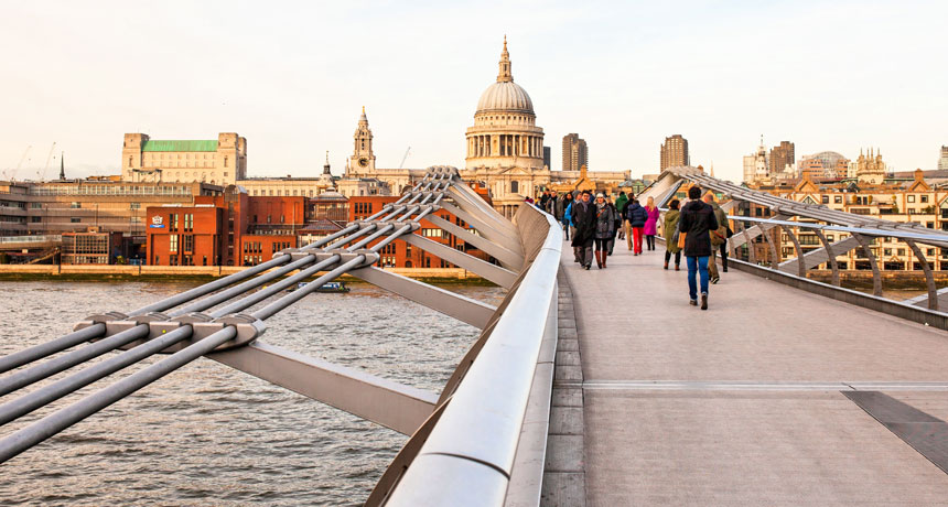
Engineers might eventually use the researchers’ results to avoid debacles like the one that befell the Millennium Bridge in London. This suspension bridge temporarily shut down just days after it opened in 2000 due to the large wobble that occurred when many people tromped across it at once (SN: 11/24/07, p. 331), necessitating costly repairs to fix the problem.
Pedestrians crossing a bridge can cause slight sideways motion of the bridge as they push with their feet. This swaying may lead to the crowd unintentionally falling into lockstep because it’s easier to go with the flow of the swinging bridge than fight it. That synchronization, in turn, creates larger and larger oscillations.
“It’s a dangerous phenomenon that could cause a bridge to collapse if it went unchecked,” says applied mathematician Daniel Abrams of Northwestern University in Evanston, Ill., who was not involved with the research.
Previous mathematical models of the phenomenon “didn’t realistically capture how people exerted force on the bridge,” Abrams says, “but this new model is pretty realistic.” Whereas earlier simulations focused on the timing of footfalls or the amount of force produced with each step, the new work takes both into account.
Tests of the Millennium Bridge showed that the lurching occurred only after a critical number of people — around 165 — entered the bridge. Likewise, in their simulations, Belykh and his colleagues find that oscillations begin abruptly above a certain threshold number of walkers, depending on the properties of the bridge.
The research challenges some previous assumptions. For instance, in the new simulations, the onset of the wobbling began just before the walkers joined in lockstep. This suggests that the synchrony of the crowd might not be a root cause but instead acts as a feedback effect that amplifies preexisting small-scale wobbles. That insight could be relevant for wobbles that occur in certain bridges without pedestrians syncing, Belykh says. Future work will further investigate how the swaying starts.