A protein made by the fetus may lead to preeclampsia in moms.
People born to mothers who had the prenatal disorder were more likely to have certain DNA variations near a gene known to influence blood vessels. The results, published online June 19 in Nature Genetics, point to that gene as a possible preeclampsia culprit, and may help scientists develop ways to stop or prevent the pregnancy complication. Preeclampsia, which is marked by a dangerous spike in blood pressure, affects about 5 percent of pregnancies and is estimated to kill over 70,000 women a year globally. Scientists have known that preeclampsia can run in families, but the genetics of the fetus hadn’t been scrutinized. “Over the years, people have looked at mothers’ genes,” says geneticist Linda Morgan of the University of Nottingham in England. “This is the first large study to look at babies’ genes.”
Morgan and colleagues compared DNA variations in 2,658 babies, children and adults born to mothers who had preeclampsia with those in more than 300,000 people. (This large group probably included some people born to mothers with the condition, but the vast majority were not.)
A genome-wide association study (GWAS), a technique used to comb through DNA looking for genetic variations that may be linked to a disorder, pinpointed a spot on chromosome 13, near a gene called FLT1. That gene is involved with blood vessel formation, an intricate process for the placenta as it grows into the inside wall of the uterus and merges the baby’s blood supply to the mother’s. The same genetic hot spot turned up in tests of a second group of offspring from mothers who had preeclampsia, Morgan and colleagues report. Another DNA variation near the gene also showed a link to the disorder.
Identifying FLT1 “makes a lot of sense,” says Ananth Karumanchi, a vascular biologist at Beth Israel Deaconess Medical Center in Boston, who was not involved in the study. Earlier experiments by Karumanchi and others suggest that the gene plays a role in preeclampsia.
Preeclampsia is kicked off by the placenta, an organ grown mostly from fetal cells that helps provide nutrients to the fetus. And though the details are unclear, some scientists suspect that unhealthy placentas start to pump out too much Flt-1 protein. A version of the protein called sFlt-1 can then slip into a mother’s bloodstream, where it may damage blood vessels in a way that leads to high blood pressure. The GWAS results can’t explain the bulk of preeclampsia cases. A fetus carrying a single copy of one of the troublesome variants near FLT1 raised a mother’s risk of preeclampsia by about 20 percent, the analysis suggests. Other risk factors are known to be much stronger, Morgan says, including previous high blood pressure, former preeclampsia diagnoses or carrying twins.
Karumanchi says that the genetic results might not be strong enough on their own to make the case that the gene is involved. But other work points to FLT1. “We feel it’s the right target,” he says.
In Europe, a preliminary clinical trial is testing a filtration method that removes excess sFlt-1 protein from the blood of women with signs of preeclampsia. So far, about 20 women have undergone the procedure, says nephrologist Ravi Thadhani of Massachusetts General Hospital in Boston. Early results are “quite encouraging,” he says, and he hopes to expand the study soon.
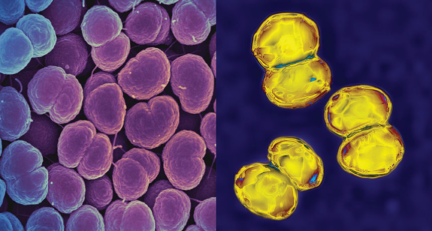
A vaccine against meningitis has an unexpected side effect: It appears to target gonorrhea, too. If confirmed, the results represent the first instance of a vaccine reducing gonorrhea infections.
After receiving a vaccine aimed at a type of meningitis, people were less likely to contract gonorrhea, scientists report online June 10 in the Lancet. That’s a big deal because worldwide each year, an estimated 78 million people contract gonorrhea, a sexually transmitted disease that can cause pelvic inflammation, infertility and throat infections. Gonorrhea’s bacterial culprit, Neisseria gonorrhoeae, has developed resistance to many antibiotics, making treatment much more difficult. Some strains of gonorrhea can now resist all known antibiotics, the Word Health Organization announced July 7. “We are in desperate need for new therapies,” says Christine Johnston, an infectious disease specialist at the University of Washington in Seattle. Attempts to make a gonorrhea vaccine have failed so far. The new results are “the first to show that vaccination against gonorrhea could be possible,” Johnston says.
Finding the link between the two diseases was partly “a story of serendipity,” says study coauthor Helen Petousis-Harris, a vaccinologist at the University of Auckland in New Zealand. She and others had noted curious declines in gonorrhea cases in New Zealand, Cuba and, to a lesser extent, Norway after people had been vaccinated against a group B meningococcal bacterium, a pathogen that can cause meningitis and blood infections.
Bacteria that cause meningitis and gonorrhea are actually close kin, sharing 80 to 90 percent of their DNA. “There was certainly biological plausibility, but we needed some proof” that the vaccine really did curb gonorrhea, Petousis-Harris says.
She and colleagues looked at data from the New Zealand national vaccine registry to see who received a meningococcal vaccine that was available from 2004 to 2008, called MeNZB. That vaccination information was combined with data on over 14,000 15- to 30-year-olds who had either gonorrhea, chlamydia or both in New Zealand between 2004 and 2016.
Compared with unvaccinated people, those who had received the vaccine were about a third less likely to contract gonorrhea, the researchers found. The researchers had no information about people’s exposure to gonorrhea, only whether people were treated for the infection at a clinic. No such link was found between the vaccine and chlamydia. MeNZB is a type of vaccine called an outer membrane vesicle vaccine. By mimicking bacterial bits released as the bugs proliferate, the vaccine trains the immune system to recognize and attack the bacteria. That exact vaccine is no longer in use, but similar vaccines exist, including Bexsero, which was used to treat a meningitis B outbreak at Princeton University in 2013.
The researchers don’t yet know what part of the MeNZB vaccine may be protective against gonorrhea. “We need to understand what was magical about this vaccine,” Petousis-Harris says. That knowledge could help researchers design a more targeted gonorrhea vaccine. Other meningitis vaccines ought to be scrutinized, too, Petousis-Harris says. “It might be that we’ve got a vaccine out there that could make a significant difference.”
Novartis, the health care company that developed Bexsero, provided funds for the study, but had no input on the design or results, Petousis-Harris says. A different company, GlaxoSmithKline, has since bought Novartis’ vaccine division.
Any new treatment for gonorrhea will eventually spur the bacteria to develop resistance, says Teodora Wi, a medical officer at WHO’s Department of Reproductive Health and Research in Geneva. But a vaccine couldn’t be evaded so easily. The current result “provides a very important breakthrough in the development of gonorrhea vaccines,” she says.
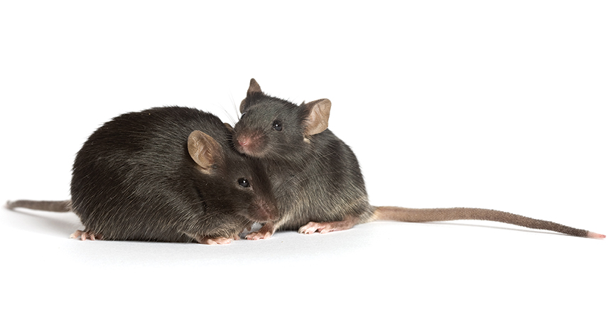
[Millions of diabetics] could be indebted to a strain of diabetic mice being bred in Bar Harbor, Maine. In diabetes research, “this mouse is the best working model to date,” one of its discoverers, Dr. Katharine P. Hummel, says.… A satisfactory animal subject had eluded diabetes researchers, until the mouse was found. — Science News, August 12, 1967
Update Hummel’s diabetic mice are still used in research to mimic type 2 diabetes in humans, which is linked to obesity. In the mid-1990s, researchers found that the diabetic mice carry a mutation in the leptin receptor gene, which prevents the hormone leptin from signaling fullness and triggering other metabolic processes. In people, however, the disease is more complicated. More than 40 genetic variants are associated with susceptibility to type 2 diabetes. Unlike the mouse mutation, none of those variants guarantee a person will develop the disease.
Across Europe, rivers aren’t flooding when they used to.
Long-term changes in temperature and precipitation are making some rivers flood days, weeks or even months earlier than they did 50 years ago, and pushing flooding in other areas much later, researchers report August 11 in Science. Those changes could impact people, wildlife and farms near rivers.
Previous studies have shown that climate change is likely to increase the severity and frequency of coastal floods, but it can be tricky to concretely link river flooding to climate change, says Günter Blöschl, a hydrologist at the Vienna University of Technology who led the study. Coastal flooding is worsened largely by one overriding variable that can be tracked: sea level rise. But river flooding is affected by a complex set of factors, says Rob Moore, a policy analyst at the Natural Resources Defense Council in Chicago who specializes in water issues. Both the timing and quantity of precipitation matter, as does the type of soil and whether it’s dry or already waterlogged when rain hits. What’s more, changes in land use around a river or engineering projects such as dams that change river flow can also affect flood risk — but aren’t necessarily related to the climate. So instead of tracking the size or frequency of river floods, the researchers examined the seasonal timing of those floods. That measurement is less impacted by factors that have nothing to do with climate. Blöschl worked with researchers from 38 countries to analyze hydrological data collected at 4,262 sites across Europe from 1960 to 2010.
Flood season shifted as much as 13 days earlier or nine days later per decade, the researchers found. Over the entire study period, that shift added up to floods in some regions occurring, in the most extreme cases, as much as 65 days earlier or 45 days later. The biggest changes were in Western Europe, where a quarter of the monitoring sites recorded flood timing shifts of more than 36 days over the 50-year period. Elsewhere, effects were more moderate, though still measurable: In northeastern Europe and the area around the North Sea, for instance, more than half of the stations showed shifts of more than 8 days. The effect varies substantially by region because not all parts of Europe experience the same sorts of floods, says Blöschl. In southern Sweden and the Baltics, floods are mostly driven by snowmelt. Warmer local temperatures make the snow melt earlier in the spring, shifting flood season up, too. In southern England, on the other hand, heavy autumn rains saturate the soil, and subsequent winter deluges can cause flooding. Flood season there is driven by when the soil gets too waterlogged to take in more moisture.
The study shows that flood timing has changed, but does not address specific consequences. It’s clear, though, that off-season flooding could have far-reaching effects, especially if these trends continue. Animals that rely on river conditions at a certain time of year in order to breed or find food could be affected by surprise floods. Out-of-season floods or unexpected dry spells could damage crops.
Plus, people are less prepared when big floods happen off-season, says Moore. While a comprehensive study like this one hasn’t been done in the United States, floods are occurring at unusual times here, too, he notes. Moore cites devastating floods that swelled the upper Mississippi River to a record size in December 2015 — not the time of year when the river is expected to overflow its banks. That flooding, combined with tornadoes spurred by the same storm system, killed more than 50 people and caused almost $2 billion in damage.
Just a stab in the dark, but you’ve probably heard: There is a total solar eclipse today, August 21.
For the first time since 1979, the moon’s shadow will zip across the continental United States. The shadow will travel from Oregon to South Carolina in a swift 92 minutes. For those in the path of totality, total darkness will last only a couple of minutes. There and elsewhere in most of the United States, the moon will partially block the sun for around three hours. If you don’t already have plans to travel to the 115-kilometer-or-so-wide path of totality, well, you’re probably too late. But here are some links to help you experience the eclipse, whether or not you’re able to see it in person.
The eclipse will be visible in all of North America — as well as in Central America and a small part of South America. Wondering what you’ll see where you live? Check out this interactive map from NASA or this cool tool from Vox.
Still need eclipse glasses? While many retailers have been sold out for days, some organizations are handing out free glasses at eclipse-watching events. Check your local TV/newspaper/radio stations’ newsfeeds for the latest. Make sure your glasses are safe.
No eclipse glasses? Never fear! You can still see the moon eclipsing the sun by making a pinhole projector or a box projector. Or just let sunlight shine through something that has holes, like a colander or Ritz Cracker (look at the ground to see the shape of the shadow the holes cast).
Watching with kids? Check out Growth Curve blogger Laura Sanders’ tips for protecting little ones’ eyes during the eclipse. Which reminds me: Whatever you do, don’t look directly at the sun. Permanent damage to your eyes may result. If you’re in the path of totality, officials say it’s OK to look directly at the sun once the moon completely blocks it. But that’s very brief, so be prepared to quickly look away or shield your eyes once the moon slips out of total alignment.
Want to do more with your eclipse experience? It’s not too late to participate in a citizen science project.
Stuck indoors, or out of totality? Watch the livestream. NASA’s programming begins at noon Eastern on NASA TV, which you can watch at this link or right here: Want some tunes to go along with it? The NASA interns made an eclipse playlist. There are also several Spotify playlists around, like this one from WXPN, this from the Washington Post and this one from the Boston Globe.
If all this excitement has you fancying a future in eclipse chasing, check out our interactive map of the next 15 total solar eclipses.
And let’s not forget that there will be a ton of science going on during the eclipse. Here are the big questions physicists and astronomers will seek to answer today.
Still want more? Follow us on Facebook and on Twitter for eclipse updates and RT’s of our correspondents in totality. Watch as the Science News team takes over the Society for Science & the Public’s Snapchat (Society4Science). And come back to Science News later today for a report from our astronomy writer, Lisa Grossman, who is spending the day in Casper, Wyo., with a research team that’s studying the sun’s wispy atmosphere, the corona.
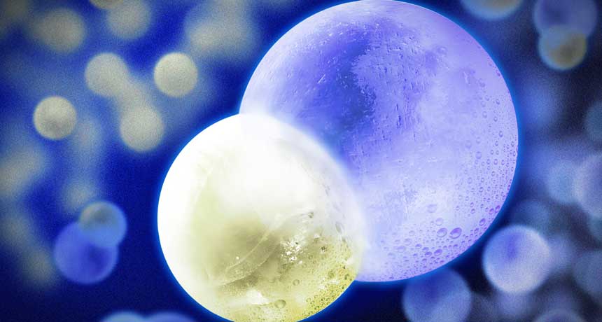
Molecules are seriously chilling out. Scientists report the first cooling of molecules below a previously impassable milestone. The result, in which scientists cooled molecules down to tens of millionths of a degree, is a step toward reaching the ultracold temperatures already achievable with atoms, researchers report August 28 in Nature Physics.
Scientists regularly chill atoms to less than a millionth of a degree above absolute zero (‒273.15° Celsius), even reaching temperatures as low as 50 trillionths of a degree (SN: 5/16/15, p. 4). But molecules are more difficult to cool down, as they can spin and vibrate in a variety of ways, and that motion is a form of heat. Previously, physicists have made ultracold molecules by convincing prechilled atoms to link up (SN: 12/20/08, p. 22), but the technique works for only a few kinds of molecules. Putting the freeze on already assembled molecules has allowed scientists to chill additional types but, until now, down to only a few hundreds of millionths of degrees.
Using lasers and magnetic fields, the scientists corralled and cooled molecules inside a device called a magneto-optical trap. In the trap, molecules of calcium monofluoride are slowed — and therefore cooled — when they absorb photons from a laser. But only so much cooling is possible with this method. To go beyond what’s called the Doppler limit, the researchers adapted a method used for cooling atoms, known as Sisyphus cooling. Two lasers pointed at one another create an electromagnetic field that acts like an endless hill the molecule must climb, thereby sapping its energy and heat. With these two techniques, the molecules reached a frigid 50 millionths of a degree above absolute zero. As the art of laser cooling advanced in recent decades, ultracold atoms rapidly became a popular research topic. Now, predicts study coauthor Michael Tarbutt, a physicist at Imperial College London, cold molecule research is “going to explode in exactly the same way that it did for cold atoms.” Cold molecules could be useful for a variety of scientific purposes: studying how chemical reactions occur, looking for hints of new fundamental particles or simulating complex quantum materials in which many particles interact at once.
“It’s a really exciting result,” says physicist David DeMille of Yale University, who was not involved with the research. “It turns out it’s harder in almost every way to apply laser cooling and trapping to molecules, but there are many, many motivations for doing that.”
Immune cells can turn certain invaders on themselves, forcing them to prematurely self-destruct, researchers have discovered.
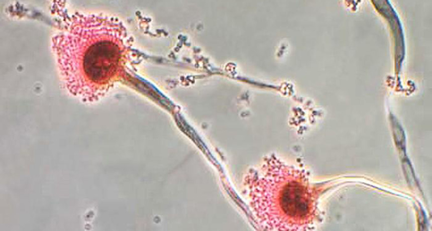
In mice, when white blood cells in the lungs engulf spores of a common airborne fungus, these immune cells release an enzyme that sends the fungal cells into programmed cell death. That prevents the spores from setting up shop in the lungs and sparking a potentially deadly lung infection, the researchers report in the Sept. 8 Science.
Found naturally in soil and decaying organic matter, the fungus, Aspergillus fumigatus, releases airborne spores that are found in small doses in the air people breathe every day. The finding may help explain why most people can regularly inhale the spores and not get sick. In people with weakened immune systems, though, this natural defense system doesn’t work. This research could eventually lead to better treatments for these patients. Programmed cell death is a natural part of a cell’s life cycle — a way for organisms to break down old cells and make way for new ones. “Research in the last couple of decades has shown that microbes can exploit [cell death] pathways to cause disease,” says study coauthor Tobias Hohl, an infectious disease researcher at Memorial Sloan Kettering Cancer Center in New York City. But this study shows that the tables can be turned. “Not only can microbes exploit this in hosts, but host cells can exploit these pathways to instruct certain microbes to kill themselves.”
“The idea that the host triggers the mechanism of [programmed cell death] as a way of defending against infection is very cool,” says Borna Mehrad, a pulmonologist at the University of Florida College of Medicine in Gainesville who wasn’t part of the study.
Hohl and colleagues identified a gene in A. fumigatus that puts the brakes on programmed cell death. The gene, AfBIR1, shares an ancestor with the human gene survivin, which also regulates cell death.
When the researchers amped up the activity of AfBIR1 in a strain of the fungus, half the mice infected with the spores died during the eight-day study period. (Mice infected with unmodified spores were fine.) Cues that would normally send fungal cells to their death didn’t register, so the fungus was able to grow in the mice’s lungs.
In another experiment, the scientists gave mice a drug called S12, which took away AfBIR1’s brake effect. As a result, the mice were able to fight off the infection. “Those two findings suggested to us that this fungal [cell death] pathway really is critical,” Hohl says. Hohl did this research with a special variety of A. fumigatus that changes color when its suicide instructions kick in. That advance allowed the researchers to make observations that weren’t possible before, Mehrad says.
For instance, Hohl and his colleagues noticed that fungal cells being engulfed by neutrophils, a type of white blood cell, appeared to be undergoing programmed cell death. That suggested that neutrophil activity might set off fungal programmed cell death.
Neutrophils release an enzyme called NADPH oxidase, and mice deficient in the enzyme weren’t as good at fending off the fungus, Hohl found. That makes sense with clinical data in humans too. People with a genetic mutation that causes a deficiency in NADPH oxidase are particularly at risk for developing an Aspergillus infection, Hohl says. People who have fewer neutrophils, due to chemotherapy or HIV infection, for instance, also make less of the enzyme and are less able to resist a fungal infection.
Survival rates vary, but the U.S. Centers for Disease Control and Prevention estimates that 41 percent of organ transplant recipients who contract aspergillosis die within a year. Seventy-five percent of stem cell transplant recipients with the infection die in that same time frame. Someday, a version of S12 that’s modified to work in humans might be able to boost these patients’ defenses against A. fumigatus infections, Hohl suggests.
In the future, he wants to see whether the same mechanisms extend to other fungal species too.
Finding a great glue is a sticky task — especially if you want it to attach to something as slick as the inside of the human body. Even the strongest human-made adhesives don’t work well on wet surfaces like tissues and organs. For surgeons closing internal incisions, that’s more than an annoyance. The right glue could hold wounds together as effectively as stitches and staples with less damage to the surrounding soft tissue, enabling safer surgical procedures.
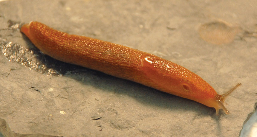
A solution might be found under wet leaves on a forest floor, recent research suggests. Jianyu Li of McGill University in Montreal and colleagues have created a surgical glue that mimics the chemical recipe of goopy slime that slugs exude when they’re startled. The adhesive stuck to a pig heart even when the surface was coated in blood, the team reported in the July 28 Science. Using the glue to plug a hole in the pig heart worked so well that the heart still held in liquid after being inflated and deflated tens of thousands of times. Li, who did the research while at Harvard University, and colleagues also tested the glue in live rats with liver lacerations. It stopped the rats’ bleeding, and the animals didn’t appear to suffer any bad reaction from the adhesive. The glue has “excellent, excellent properties,” says Andrew Smith, a biologist at Ithaca College in New York. And slugs aren’t the only biological inspiration for new adhesives. Clues to better glues have long been hiding out in damp, soggy and downright wet places. For slugs, mussels, marine worms and a cadre of other critters, secreting sticky substances that attach strongly to soaked surfaces is just a fact of life. That’s why scientists are studying the structures of those substances to design new and better surgical adhesives.
“There’s really a big need to develop new ways of sealing tissues, of affixing devices to tissues — in particular, for minimally invasive procedures,” says Jeff Karp, a biomedical engineer at Brigham and Women’s Hospital in Boston. While existing medical-grade superglue is great at sealing up fingertip cuts, it is too toxic to use inside the body. Other alternatives just aren’t sticky enough to fully replace stitches. With a better glue, surgeons could also make snips that are too tiny to be stitched or stapled closed. Smaller incisions speed healing time and decrease risk of complications, Karp says.
Smith says he isn’t surprised that slug slime could lead to a big advance. For several years, he’s been trying to understand how the slug Arion subfuscus builds its ooze. For his research, Smith prods slugs gently with the tip of a metal spatula to startle them, and scoops up the slime as it’s released. “If you get it on your hands, it’ll set within seconds into an extremely sticky material,” he says. The goo, Smith and others have found, overcomes a major challenge that adhesive designers face. It seems obvious that glue should be sticky. Yet the molecules in glue need to adhere not just to the things you’re trying to stick together, but also to each other. And that stickiness can’t come at the expense of flexibility, especially for medical applications. Soft, squishy organs are going to jiggle; skin is going to stretch. Without some bendiness, the glue might attach securely to each of the surfaces being stuck together, but the glob of glue itself might snap or shear under stress.
Slug defense slime solves that problem with two interwoven networks of molecules, tangled together like strings of holiday lights. One network is rigid, with chemical bonds that break easily, Smith says. The other is deformable, stretching substantially without breaking. This combo makes the goo simultaneously tough, flexible and sticky.
Li’s slug-inspired adhesive takes a similar approach. One layer of the material is a polymer, a type of material made from long molecules built from many repeated subunits, like a string of beads. Positively charged appendages dangling off the polymers are drawn to wet tissue surfaces by the same forces underlying static electricity. This first layer weaves into another layer, a water-based gel. The gel layer acts like a shock absorber in a car, Li says. It soaks up energy that might otherwise dislodge or snap the adhesive.
Despite being 90 percent water, the material is both sticky and tough, Li says. The fact that it’s mostly water makes it more likely to be nontoxic to humans. Though Li’s adhesive has been tested only in human cell cultures and in lab animals, another bio-inspired glue has made its way into human trials. It’s based on work published by Karp and colleagues in 2014 in Science Translational Medicine. Karp’s team developed a viscous liquid that solidifies into a tough but stretchy glue when illuminated by light, and demonstrated that the liquid can seal holes in hearts.
“Nothing we create is really that similar to anything you see in nature, but some of the ideas gave us critical insights,” Karp says. The researchers realized, for example, that a lot of natural glues that work in water have hydrophobic elements that help clear away the water for a better stick. The research sparked Karp and colleagues to found a company, Gecko Biomedical, which Karp now advises. On September 11, the company announced the completion of a small clinical trial of its adhesive: The sealant immediately stopped blood flow after an artery-clearing operation in about 85 percent of 22 participants. Because of that success, Gecko Biomedical now has approval to market the glue in Europe.
Bio-inspired adhesives can do more than patch up incisions, though. Russell Stewart, a bioengineer at the University of Utah in Salt Lake City, is tapping into marine-dwelling sandcastle worms for a different glue goal: He wants to create a better embolic agent — a way to deliberately block blood flow to certain tissues. Embolic agents can cut blood flow to a tumor, say, or stem internal bleeding. Often, these materials are liquids that reach their target through a catheter and then solidify into a sticky mass to block tiny vessels. But such glues can be difficult to control — they need to harden at just the right time and current options often rely on harsh materials that require special equipment and can cause pain for patients.
Inspired by the sandcastle worm (Phragmatopoma californica), Stewart has designed a new — and he thinks better — embolic agent. A sandcastle worm uses fingerlike appendages coming out of its face to arrange grains of sand into expansive tubular reefs. It squirts small dabs of a liquid adhesive out of these appendages to make the grains stick together. That glue’s structure is quite different from slug slime, Stewart has found. It’s a solution of oppositely charged proteins strongly attracted to each other. The proteins make up a dense liquid that doesn’t mix with water. A worm packages each ingredient in the glue separately, so the proteins combine only once secreted. After mixing, the glue solidifies in about 30 seconds.
Stewart’s mimic also starts out as a liquid that transforms into a hard foamlike material within a few seconds of hitting blood, his team reported in 2016 in Advanced Healthcare Materials. That means the material can be injected as a liquid and doesn’t harden until it’s in the right place. Early tests have been promising: The foam completely blocked the arteries of rabbits’ kidneys without moving into tissue where it didn’t belong.
The range of biological adhesives is impressive, says Jonathan Wilker, a chemist at Purdue University in West LaFayette, Ind. “They’re so wildly different,” both in terms of chemical makeup and functional properties. That diversity provides a wide palette for scientists seeking glues for specialized applications. And Wilker’s own work adds mussels to the list.
Mussels secrete a strong adhesive that helps them stick tenaciously to rocks and ship hulls. Their secret is a molecule called DOPA, Wilker says. DOPA, or 3,4-dihydroxyphenylalanine, sticks well to other DOPA molecules and to other substances. That gives it the same balance of toughness and stickiness that’s also found in slug slime. Certain amino acids found in mussel proteins might also aid the underwater adhesion. For example, an amino acid called lysine that hangs off of mussel adhesion proteins appears to help clear water molecules out of the way, leaving a drier surface for proteins glomming on. Wilker’s copycat adhesive is made up of long chains of polystyrene molecules (essentially, Styrofoam) with units of DOPA mixed in. Those long chains of tricked-out polystyrene molecules tangle together and cross-link to create a strong adhesive. He’s made different varieties of the mimic, tailored for different applications. After being immersed in water, one version held on tighter underwater than the glue made by mussels themselves, Wilker’s team reported in February in Applied Materials Interfaces. Another version is biodegradable.
If he can make the glues nontoxic to cells, they could possibly be used inside the body. In one recent study, Wilker created an artificial adhesive protein that mimics the natural protein elastin. The artificial version excelled in both dry and damp test environments, his team reported in April in Biomaterials.
Bringing animal-inspired adhesives into the human body won’t necessarily be a simple task, though. It requires tackling some problems that other animals don’t need to solve, Karp says. A slug, for instance, produces its slime as it needs it. It doesn’t stockpile gallons of glue in its tiny body, or instantly churn out a year’s supply. A successful real-world glue, however, will need to be easy to produce in large quantities and safe to store for months at a time, Karp points out. Those are problems humans will have to solve on their own. That’s the next challenge.
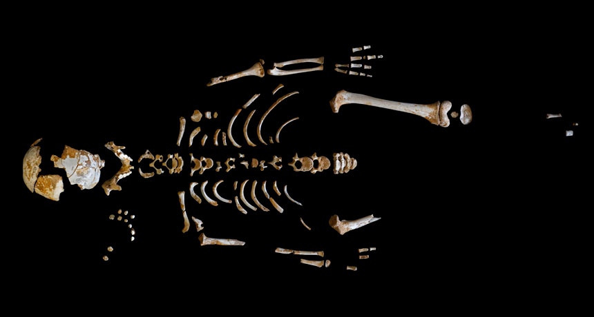
A Neandertal child whose partial skeleton dates to around 49,000 years ago grew at the same pace as children do today, with a couple of exceptions. Growth of the child’s spine and brain lagged, a new study finds.
It’s unclear, though, whether developmental slowing in those parts of the body applied only to Neandertals or to Stone Age Homo sapiens as well. If so, environmental conditions at the time — which are currently hard to specify — may have reduced the pace of physical development similarly in both Homo species. This ancient youngster died at 7.7 years of age, say paleoanthropologist Antonio Rosas of the National Museum of Natural Sciences in Madrid and colleagues. The scientists estimated the child’s age by counting microscopic enamel layers that accumulated daily as a molar tooth formed.
Previous excavations uncovered the child’s remains, as well as fossils of 12 other Neandertals, at a cave site in northwestern Spain called El Sidrόn.
Much — but not all —of the Neandertal child’s skeleton had matured to a point expected for present-day youngsters of the same age, the scientists report in the Sept. 22 Science. But bones at the top and in the middle of the spine had not fully fused, corresponding to a stage of development typical of 4- to 6- year-olds today. Also, the ancient child’s brain was still growing at an age when living humans’ brains have nearly or fully reached adult size. Signs of bone tissue being reshaped on the inner surface of the child’s braincase pointed to ongoing brain expansion. Rosas’ team calculated that the youngster’s brain volume was about 87.5 percent of that expected, on average, for Neandertal adults.
Neandertals’ slightly larger brains relative to people today may have required more energy, and thus more time, to grow, the researchers suggest. And they suspect that the growth of Neandertals’ bigger torsos, and perhaps spinal cords, slowed the extinct species’ backbone development in late childhood.
Rosas’ new study “reinforces what should have been apparent for some time — that Neandertal growth rates and patterns, except for those related to well-known differences in [skeletal shape], rarely differ from modern human variations,” says paleoanthropologist Erik Trinkaus of Washington University in St. Louis.
But researchers need to compare the El Sidrόn child to fossils of H. sapiens youngsters from the same time or later in the Stone Age, Trinkaus adds. Relative to kids today, ancient human youth may display slower growth rates comparable to those of the Neandertal child, he suspects.
Subtle cosmic vibrations kicked up by swirling black holes have captured the public imagination — and the minds of the physics Nobel Prize committee members, too.
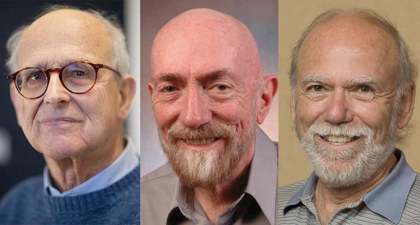
Three scientists who laid the groundwork for the first direct detection of gravitational waves have won the Nobel Prize in physics. Rainer Weiss of MIT, and Kip Thorne and Barry Barish, both of Caltech, will share the 9-million-Swedish-kronor (about $1.1 million) prize, with half going to Weiss and the remainder split between Thorne and Barish. Though researchers often wait decades for Nobel recognition, the observation of gravitational waves was so monumental that the scientists were honored less than two years after the discovery’s announcement.
“These detections were so compelling and earth shattering…. Why wait?” says Clifford Will of the University of Florida in Gainesville, who was not directly involved with the discovery. “It’s fabulous. Absolutely fabulous.”
Weiss, Thorne and Barish are pioneers of the Laser Interferometer Gravitational Wave Observatory, or LIGO. On February 11, 2016, LIGO scientists announced they had spotted gravitational waves produced by a pair of merging black holes. This first-ever detection generated a frenzy of excitement among physicists and garnered front-page headlines around the world.
LIGO’s observation of gravitational waves directly confirmed a 100-year-old prediction of Einstein’s general theory of relativity — that rapidly accelerating massive objects stretch and squeeze spacetime, producing ripples that travel outward from the source (SN: 3/5/16, p. 22). “If Einstein was still alive, it would be absolutely wonderful to go to him and tell him about the discovery. He would be very pleased, I’m sure of it,” Weiss said during a news conference at MIT a few hours after he got word of the win. “But then to tell him what the discovery was, that it was a black hole, he would have been absolutely flabbergasted because he didn’t believe in them.”
As enthusiastic team members clad in LIGO-themed T-shirts celebrated the discovery, Weiss stressed that the discovery was a group effort. “I’m a symbol of that. It’s not all on my shoulders, this thing,” he said, citing the large collaboration of scientists whose work led up to LIGO’s detection.
Physicists anticipate that LIGO will spark an entirely new field of astronomy, in which scientists survey the universe by feeling for its tremors. “It will allow us to see the parts of the universe that were not revealed to us before,” says LIGO team member Carlos Lousto of the Rochester Institute of Technology in New York.
LIGO’s first incarnation, which officially began collecting data in 2002 and ran intermittently until 2010, yielded no hints of gravitational waves. After years of upgrades, the souped-up detectors, known as Advanced LIGO, began searching for spacetime ripples in 2015. Almost as soon as the detectors were turned on — even before scientific data-taking had formally begun — scientists detected the minuscule undulations of their first black hole collision. Those ripples, spotted on September 14, 2015, journeyed to Earth from 1.3 billion light-years away, where they were produced by two colossal black holes that spiraled inward and merged into one (SN: 3/5/16, p. 6).
Quivers from those converging black holes, when converted into an audio signal, made a tell-tale sound called a “chirp,” reminiscent of a bird’s cry. The particulars of that signature reveal details of the collision. “The beauty of the symphony is in what you can extract from the tiny wiggles, or the wiggles on tops of wiggles, in that signal,” Thorne said at an Oct. 3 news conference at Caltech. Since that first detection, scientists have observed three more black hole collisions. And additional gravitational ripples may already be in the bag: It’s rumored that LIGO scientists have also detected a smashup of neutron stars (SN Online: 8/25/17). In fact, Weiss teased an announcement to come on October 16.
An astounding feat of engineering, LIGO consists of two enormous L-shaped detectors that stretch across the wooded landscape of Livingston, La., and the desert of Hanford, Wash. Each detector boasts two 4-kilometer-long arms through which laser light bounces back and forth between mirrors.
Gravitational waves passing through a detector stretch one arm while shortening the other. LIGO compares the arms’ sizes using the laser light to measure length differences a tiny fraction of the size of a proton. Gravitational waves should produce signals in the two distant detectors nearly simultaneously, helping scientists to rule out spurious signals that can be caused by events as mundane as a truck bouncing along nearby.
“LIGO is probably one of the best and most amazing instruments ever built by mankind,” Barish said at the Caltech news conference. But building it was a risky endeavor: No one had previously attempted anything like it, and no one could say for sure whether the effort would succeed. “What’s fundamental is you have to be willing to take risks to do great things,” Barish said.
In August, LIGO’s two detectors teamed up with the similarly designed Virgo detector near Pisa, Italy (SN Online: 8/1/17). The latest gravitational wave sighting, made on August 14, showed up in all three detectors almost simultaneously, which allowed scientists to pinpoint the region of space in which the black holes resided more precisely than ever before (SN Online: 9/27/17).
Weiss spent decades on the project, beginning with nascent scribbles on scraps of paper and early prototypes. In the 1960s, Weiss came up with the idea for a laser gravitational wave detector while teaching a class on general relativity. (Other researchers had independently proposed the technique as well.) He refined that idea and built a small, prototype detector, establishing the basic blueprint that would eventually evolve into LIGO. Inspired by a conversation with Weiss, Thorne, who had been studying theoretical aspects of gravitational waves, assembled a team to work on the technique at Caltech in the ’70s. (Thorne was a 1958 semifinalist in the Science Talent Search, a program of the Society for Science & the Public, which publishes Science News.)
Another LIGO founder, Ronald Drever, died in March. Drever, who had been working on gravitational wave detectors at the University of Glasgow, joined Thorne at Caltech in 1979. Weiss and Drever each worked individually on prototypes, before Weiss officially teamed up with Thorne and Drever in 1984 to create LIGO (SN: 3/5/16, p. 24). Drever did live to hear of the first detection, Will says, but “it’s sad that he didn’t live to see it all.”
Barish joined the project later, becoming director of LIGO in 1994. He stayed in that role for more than 10 years, elevating LIGO from scientists’ daydreams into reality. Barish oversaw construction and commissioning of the detectors, as well as initial gravitational wave searches. “He entered the experiment in a crucial moment, when it was necessary to bring the experiment to a different level, make it a big collaboration,” says Alessandra Buonanno of the Max Planck Institute for Gravitational Physics in Potsdam, Germany.
Speculation that LIGO would nab a Nobel began as soon as the discovery was announced. So the collaboration was not surprised by the honor. “We were certainly expecting this to happen,” says LIGO team member Manuela Campanelli of the Rochester Institute of Technology. Still, the lack of surprise didn’t dampen the mood of festivity. “I feel in a dream,” says Buonanno.
LIGO and Virgo are currently in a shutdown period while scientists tinker with the detectors to improve their sensitivity. The gravitational wave hunt will resume next year. Besides black hole mergers and neutron star smashups, in the future, scientists might also spot waves from an exploding star, known as a supernova. Upcoming detectors might sense trembles generated in the Big Bang, providing a glimpse of the universe’s beginnings.
And scientists may even find new phenomena that they haven’t predicted. “I await expectantly some huge surprises in the coming years,” Thorne said.